Cellular and Molecular Imaging Laboratory, Department of Radiology, Henry Ford Hospital, Detroit, Michigan, United States of America.
PLoS One. 2010 Feb 26;5(2):e9365. doi: 10.1371/journal.pone.0009365.
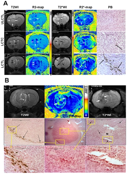
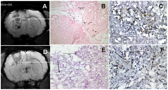
A limitation with current imaging strategies of recurrent glioma undergoing radiotherapy is that tumor and radiation injury cannot be differentiated with post contrast CT or MRI, or with PET or other more complex parametric analyses of MRI data. We propose to address the imaging limitation building on emerging evidence indicating that effective therapy for recurrent glioma can be attained by sensitized T-cells following vaccination of primed dendritic cells (DCs). The purpose of this study was to determine whether cord blood T-cells can be sensitized against glioma cells (U-251) and if these sensitized cytotoxic T-cells (CTLs) can be used as cellular magnetic resonance imaging probes to identify and differentiate glioma from radiation necrosis in rodent models.
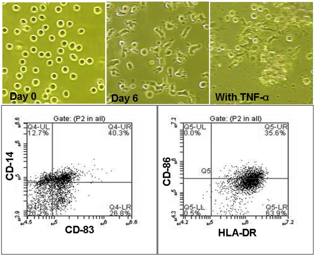
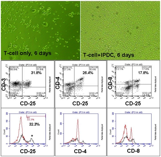
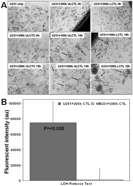
METHODOLOGY/PRINCIPAL FINDINGS: Cord blood T and CD14+ cells were collected. Isolated CD14+ cells were then converted to dendritic cells (DCs), primed with glioma cell lysate and used to sensitize T-cells. Phenotypical expression of the generated DCs were analyzed to determine the expression level of CD14, CD86, CD83 and HLA-DR. Cells positive for CD25, CD4, CD8 were determined in generated CTLs. Specificity of cytotoxicity of the generated CTLs was also determined by lactate dehydrogenase (LDH) release assay. Secondary proliferation capacity of magnetically labeled and unlabeled CTLs was also determined. Generated CTLs were magnetically labeled and intravenously injected into glioma bearing animals that underwent MRI on days 3 and 7 post- injection. CTLs were also administered to animals with focal radiation injury to determine whether these CTLs accumulated non-specifically to the injury sites. Multi-echo T2- and T2*-weighted images were acquired and R2 and R2* maps created. Our method produced functional, sensitized CTLs that specifically induced U251 cell death in vitro. Both labeled and unlabeled CTLs proliferated equally after the secondary stimulation. There were significantly higher CD25 positive cells (p = <0.006) in CTLs. In addition, T2- and T2*-weighted MR images showed increased low signal intensity areas in animals that received labeled CTLs as compared to the images from animals that received control cells. Histological analysis confirmed the presence of iron positive cells in sites corresponding to MRI low signal intensity regions. Significant differences (p = <0.001) in tumor R2 and R2* values were observed among the groups of animals. Animals with radiation injury exhibited neither MRI hypointense areas nor presence of iron positive cells.
Our results indicate that T-cells can be effectively sensitized by in vitro methods and used as cellular probes to identify and differentiate glioma from radiation necrosis.
目前对接受放疗的复发性神经胶质瘤进行成像的策略存在一个局限性,即肿瘤和辐射损伤无法通过增强 CT 或 MRI,或通过 PET 或 MRI 数据的其他更复杂参数分析来区分。我们建议基于新兴证据来解决成像限制,这些证据表明,通过接种致敏树突状细胞(DC),可以使复发性神经胶质瘤患者获得有效的治疗效果。本研究的目的是确定脐带血 T 细胞是否可以针对神经胶质瘤细胞(U-251)进行致敏,以及这些致敏的细胞毒性 T 细胞(CTL)是否可以用作细胞磁共振成像探针,以在啮齿动物模型中识别和区分神经胶质瘤与放射性坏死。
方法/主要发现:收集脐带血 T 细胞和 CD14+细胞。分离出的 CD14+细胞随后被转化为树突状细胞(DC),用神经胶质瘤细胞裂解物进行致敏,并用于致敏 T 细胞。分析生成的 DC 的表型表达,以确定 CD14、CD86、CD83 和 HLA-DR 的表达水平。在生成的 CTL 中确定 CD25、CD4、CD8 阳性细胞。通过乳酸脱氢酶(LDH)释放测定也确定了生成的 CTL 的细胞毒性特异性。还确定了经磁标记和未标记 CTL 的二次增殖能力。生成的 CTL 被磁标记并静脉注射到接受放疗的神经胶质瘤荷瘤动物中,在注射后第 3 天和第 7 天进行 MRI。还将 CTL 施用于具有局灶性放射性损伤的动物,以确定这些 CTL 是否会非特异性地积聚在损伤部位。采集多回波 T2-和 T2*-加权图像,并创建 R2 和 R2图谱。我们的方法产生了功能正常的致敏 CTL,可特异性诱导 U251 细胞在体外死亡。在二次刺激后,标记和未标记的 CTL 增殖能力相同。CTL 中 CD25 阳性细胞明显更多(p = <0.006)。此外,与接受对照细胞的动物的图像相比,接受标记 CTL 的动物的 T2-和 T2-加权 MR 图像显示出低信号强度区域增加。组织学分析证实 MRI 低信号强度区域对应的部位存在铁阳性细胞。在动物组中观察到肿瘤 R2 和 R2*值的显著差异(p = <0.001)。具有放射性损伤的动物既没有 MRI 低信号强度区域,也没有铁阳性细胞。
我们的结果表明,T 细胞可以通过体外方法有效地致敏,并用作细胞探针来识别和区分神经胶质瘤与放射性坏死。