Institute of Anatomy, Kaunas University of Medicine, Kaunas, Lithuania.
Heart Rhythm. 2010 Jul;7(7):942-50. doi: 10.1016/j.hrthm.2010.02.036. Epub 2010 Mar 1.
Sheep are routinely used in experimental cardiac electrophysiology and surgery.
The purpose of this study was to (1) ascertain the topography and architecture of the ovine epicardial neural plexus (ENP), (2) determine the relationships of ENP with vagal and sympathetic cardiac nerves and ganglia, and (3) evaluate gross anatomic differences and similarities of ENP in humans, sheep, and other species.
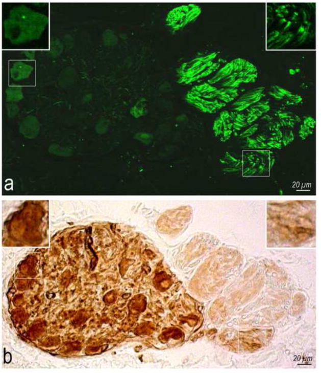
Ovine ENP and extrinsic sympathetic and vagal nerves were stained histochemically for acetylcholinesterase in whole heart and/or thorax-dissected preparations from 23 newborn lambs, with subsequent examination by stereomicroscope.
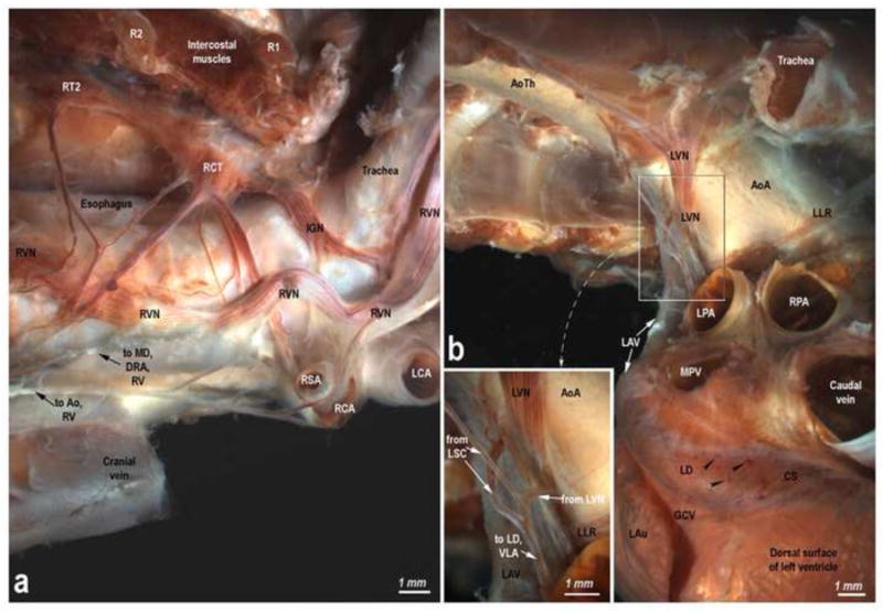
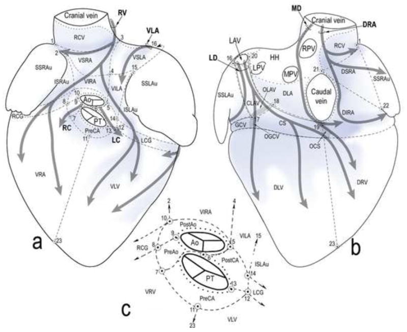
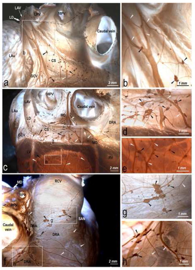
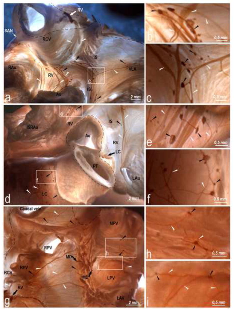
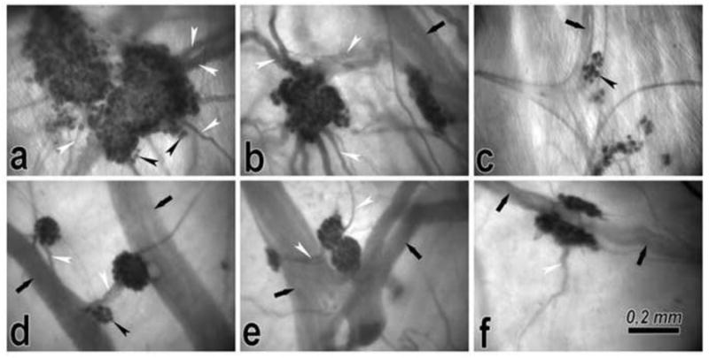
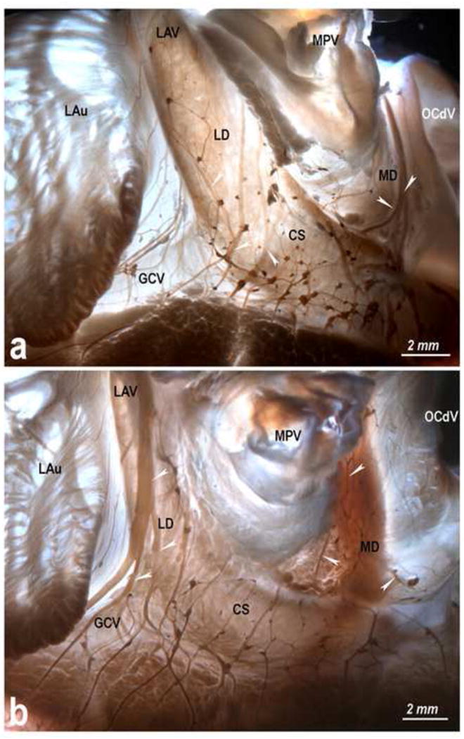
Intrinsic cardiac nerves extend from the venous part of the ovine heart hilum along the roots of the cranial (superior) caval and left azygos veins to both atria and ventricles via five epicardial routes: dorsal right atrial, middle dorsal, left dorsal, right ventral, and ventral left atrial nerve subplexuses. Intrinsic nerves proceeding from the arterial part of the heart hilum along the roots of the aorta and pulmonary trunk extend exclusively into the ventricles as the right and left coronary subplexuses. The dorsal right atrial, right ventral, and middle dorsal subplexuses receive the main extrinsic neural input from the right cervicothoracic and right thoracic sympathetic T(2) and T(3) ganglia as well as from the right vagal nerve. The left dorsal is supplied by sizeable extrinsic nerves from the left thoracic T(4)-T(6) sympathetic ganglia and the left vagal nerve. Sheep hearts contained an average of 769 +/- 52 epicardial ganglia. Cumulative areas of epicardial ganglia on the root of the cranial vena cava and on the wall of the coronary sinus were the largest of all regions (P <.05).
Despite substantial interindividual variability in the morphology of ovine ENP, right-sided epicardial neural subplexuses supplying the sinoatrial and atrioventricular nodes are mostly concentrated at a fat pad between the right pulmonary veins and the cranial vena cava. This finding is in sharp contrast with a solely left lateral neural input to the human atrioventricular node, which extends mainly from the left dorsal and middle dorsal subplexuses. The abundance of epicardial ganglia distributed widely along the ovine ventricular nerves over respectable distances below the coronary groove implies a distinctive neural control of the ventricles in human and sheep hearts.
绵羊常用于心脏电生理学和心脏手术的实验研究。
本研究的目的是:(1)确定绵羊心外膜神经丛(ENP)的拓扑结构和形态学,(2)确定 ENP 与迷走神经和交感神经心脏神经和神经节的关系,以及(3)评估人类、绵羊和其他物种的 ENP 的大体解剖差异和相似性。
对 23 只新生羔羊的整个心脏和/或胸腔解剖心脏进行乙酰胆碱酯酶的组织化学染色,以研究绵羊 ENP 以及外生交感神经和迷走神经,随后通过立体显微镜进行检查。
心脏固有神经从心尖静脉部分沿头腔静脉和左奇静脉的根部延伸至左右心房和心室,通过五条心外膜途径:右心房背侧、中背侧、左背侧、右腹侧和左腹侧心房神经亚丛。从心脏动脉部分沿主动脉和肺动脉根部延伸的固有神经仅延伸至心室,作为右冠状动脉和左冠状动脉亚丛。右心房背侧、右腹侧和中背侧亚丛主要接收来自右颈胸和右胸 T(2)和 T(3)交感神经节以及右迷走神经的外生神经输入。左背侧由来自左胸 T(4)-T(6)交感神经节和左迷走神经的大量外生神经供应。绵羊心脏平均含有 769 +/- 52 个心外膜神经节。颅腔静脉根部和冠状窦壁的心脏外膜神经节累积面积是所有区域中最大的(P <.05)。
尽管绵羊 ENP 的形态存在很大的个体间变异性,但供应窦房结和房室结的右侧心外膜神经亚丛主要集中在右肺静脉和头腔静脉之间的脂肪垫上。这一发现与人类房室结主要由左背侧和中背侧亚丛主要从左侧提供的单纯左侧神经输入形成鲜明对比。广泛分布在绵羊心室神经上的大量心外膜神经节沿着心室神经延伸可观的距离,在冠状沟下方,这意味着人类和绵羊心脏的心室有独特的神经控制。