Department of Developmental Neurobiology, NYS Institute for Basic Research in Developmental Disabilities (IBR), 1050 Forest Hill Road, Staten Island, NY 10314, USA.
Acta Neuropathol. 2010 Jun;119(6):755-70. doi: 10.1007/s00401-010-0655-4. Epub 2010 Mar 3.
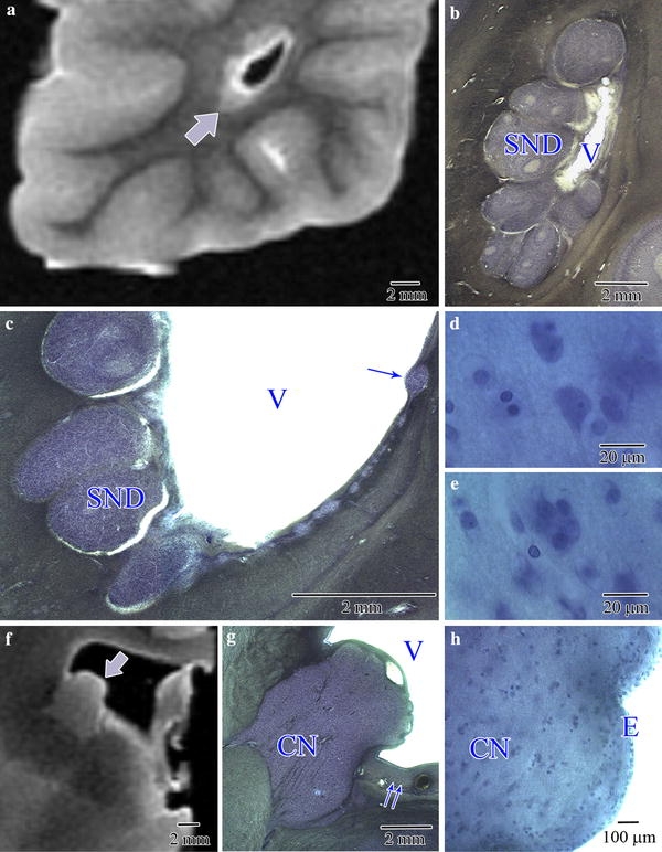
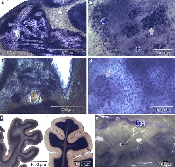
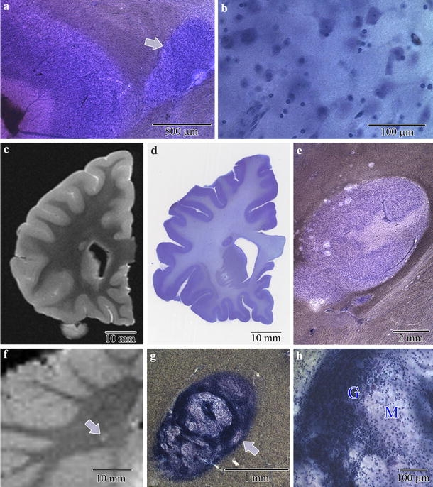
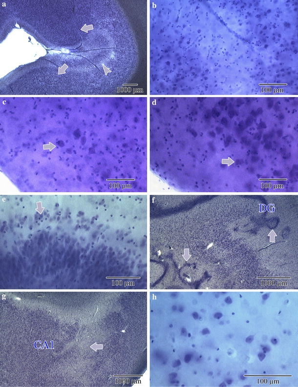
Autism is characterized by a broad spectrum of clinical manifestations including qualitative impairments in social interactions and communication, and repetitive and stereotyped patterns of behavior. Abnormal acceleration of brain growth in early childhood, signs of slower growth of neurons, and minicolumn developmental abnormalities suggest multiregional alterations. The aim of this study was to detect the patterns of focal qualitative developmental defects and to identify brain regions that are prone to developmental alterations in autism. Formalin-fixed brain hemispheres of 13 autistic (4-60 years of age) and 14 age-matched control subjects were embedded in celloidin and cut into 200-mum-thick coronal sections, which were stained with cresyl violet and used for neuropathological evaluation. Thickening of the subependymal cell layer in two brains and subependymal nodular dysplasia in one brain is indicative of active neurogenesis in two autistic children. Subcortical, periventricular, hippocampal and cerebellar heterotopias detected in the brains of four autistic subjects (31%) reflect abnormal neuronal migration. Multifocal cerebral dysplasia resulted in local distortion of the cytoarchitecture of the neocortex in four brains (31%), of the entorhinal cortex in two brains (15%), of the cornu Ammonis in four brains and of the dentate gyrus in two brains. Cerebellar flocculonodular dysplasia detected in six subjects (46%), focal dysplasia in the vermis in one case, and hypoplasia in one subject indicate local failure of cerebellar development in 62% of autistic subjects. Detection of flocculonodular dysplasia in only one control subject and of a broad spectrum of focal qualitative neuropathological developmental changes in 12 of 13 examined brains of autistic subjects (92%) reflects multiregional dysregulation of neurogenesis, neuronal migration and maturation in autism, which may contribute to the heterogeneity of the clinical phenotype.
自闭症的临床表现广泛,包括社交互动和沟通方面的质的损伤,以及重复和刻板的行为模式。儿童早期大脑生长加速异常、神经元生长速度较慢的迹象以及微小柱发育异常表明多区域改变。本研究旨在检测局灶性质的发育缺陷模式,并确定大脑区域容易发生自闭症的发育改变。将 13 名自闭症(4-60 岁)和 14 名年龄匹配的对照者的福尔马林固定的大脑半球包埋在细胞素中,并切成 200μm 厚的冠状切片,用甲苯胺蓝染色进行神经病理学评估。两名自闭症儿童的室管膜下细胞层增厚和一个大脑的室管膜下结节性发育不良提示活跃的神经发生。四名自闭症患者(31%)的大脑中发现的皮质下、脑室周围、海马和小脑异位提示异常神经元迁移。四个大脑(31%)的局部皮质结构紊乱导致多灶性脑发育不良,两个大脑(15%)的内嗅皮质,四个大脑的角回和两个大脑的齿状回。六个患者(46%)的小脑绒球小结叶发育不良,一个病例的蚓部局灶性发育不良,一个患者的小脑发育不全,表明 62%的自闭症患者的小脑发育局部失败。仅在一个对照者中检测到绒球小结叶发育不良,而在自闭症患者的 13 个检查大脑中,有 12 个(92%)表现出广泛的局灶性质的神经病理学发育变化,这反映了自闭症中神经发生、神经元迁移和成熟的多区域失调,这可能导致临床表型的异质性。