Department of General Surgery, Molecular Oncology and Immunology, University of Wuerzburg Hospital, Germany.
BMC Cancer. 2010 Mar 7;10:82. doi: 10.1186/1471-2407-10-82.
The local and systemic activation and regulation of the immune system by malignant cells during carcinogenesis is highly complex with involvement of the innate and acquired immune system. Despite the fact that malignant cells do have antigenic properties their immunogenic effects are minor suggesting tumor induced mechanisms to circumvent cancer immunosurveillance. The aim of this study is the analysis of tumor immune escape mechanisms in a colorectal liver metastases mouse model at different points in time during tumor growth.
CT26.WT murine colon carcinoma cells were injected intraportally in Balb/c mice after median laparotomy using a standardized injection technique. Metastatic tumor growth in the liver was examined by standard histological procedures at defined points in time during metastatic growth. Liver tissue with metastases was additionally analyzed for cytokines, T cell markers and Fas/Fas-L expression using immunohistochemistry, immunofluorescence and RT-PCR. Comparisons were performed by analysis of variance or paired and unpaired t test when appropriate.
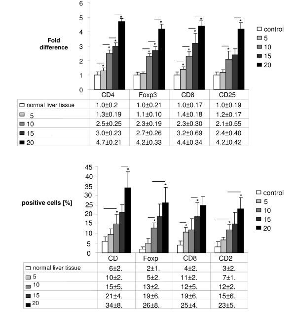
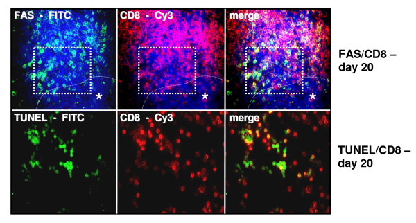
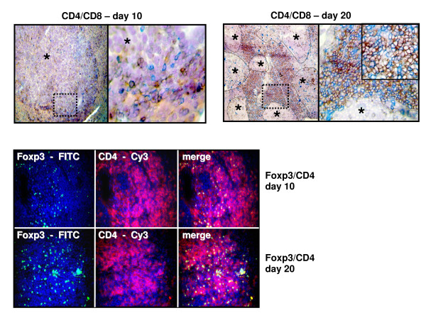
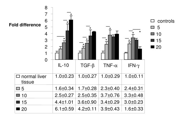
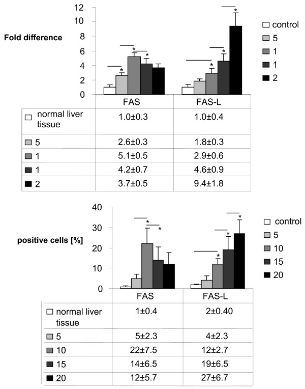
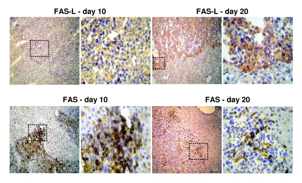
Intraportal injection of colon carcinoma cells resulted in a gradual and time dependent metastatic growth. T cells of regulatory phenotype (CD4+CD25+Foxp3+) which might play a role in protumoral immune response were found to infiltrate peritumoral tissue increasingly during carcinogenesis. Expression of cytokines IL-10, TGF-beta and TNF-alpha were increased during tumor growth whereas IFN-gamma showed a decrease of the expression from day 10 on following an initial increase. Moreover, liver metastases of murine colon carcinoma show an up-regulation of FAS-L on tumor cell surface with a decreased expression of FAS from day 10 on. CD8+ T cells express FAS and show an increased rate of apoptosis at perimetastatic location.
This study describes cellular and macromolecular changes contributing to immunological escape mechanisms during metastatic growth in a colorectal liver metastases mouse model simulating the situation in human cancer.
恶性细胞在癌变过程中通过局部和全身激活和调节免疫系统,这一过程非常复杂,涉及先天和获得性免疫系统。尽管恶性细胞确实具有抗原特性,但它们的免疫原性效应较小,这表明肿瘤诱导的机制可以规避癌症免疫监视。本研究的目的是在结直肠肝转移小鼠模型中分析肿瘤免疫逃逸机制,即在肿瘤生长的不同时间点。
采用标准注射技术,通过中腹部剖腹手术将 CT26.WT 小鼠结肠癌细胞注入 Balb/c 小鼠门静脉内。在转移性生长的特定时间点,通过标准组织学程序检查肝脏中的转移性肿瘤生长。使用免疫组织化学、免疫荧光和 RT-PCR 分析肝组织中的细胞因子、T 细胞标志物和 Fas/Fas-L 表达。通过方差分析或适当的配对和非配对 t 检验进行比较。
门静脉内注射结肠癌细胞导致逐渐和时间依赖性的转移性生长。在癌变过程中,调节表型的 T 细胞(CD4+CD25+Foxp3+)逐渐浸润肿瘤周围组织,这些 T 细胞可能在促进肿瘤免疫反应中发挥作用。细胞因子 IL-10、TGF-β 和 TNF-α 的表达在肿瘤生长过程中增加,而 IFN-γ 的表达则在最初增加后从第 10 天开始下降。此外,小鼠结肠癌细胞的肝转移显示肿瘤细胞表面 Fas-L 的上调和 Fas 的表达从第 10 天开始下降。CD8+T 细胞表达 Fas,并在肿瘤周围位置表现出更高的凋亡率。
本研究描述了在结直肠肝转移小鼠模型中,在模拟人类癌症的情况下,导致转移性生长过程中免疫逃逸机制的细胞和大分子变化。