Department of Surgery, University of Illinois at Chicago, Division of Surgical Oncology, 835 S, Wolcott MC790, Chicago, IL 60612, USA.
J Transl Med. 2013 Mar 20;11:70. doi: 10.1186/1479-5876-11-70.
T-cell infiltration in primary colon tumors is associated with improved patient survival. Preliminary data supports a similar association in colorectal liver metastases (CRLM), and we previously identified increased CRLM expression of the immunostimulatory cytokine LIGHT (TNFSF14) to be related to improved patient prognosis. Therefore, mechanisms to augment the T-cell response in CRLM may be a promising treatment modality, however, the tumor immune microenvironment and LIGHT expression in CRLM remains to be characterized.
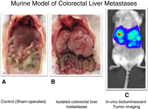
Utilizing a syngeneic and immunocompetent model of CRLM, the immune microenvironment was characterized for lymphocyte phenotype, function, and location utilizing flow cytometry, immunoassays, and immunofluorescence microscopy.
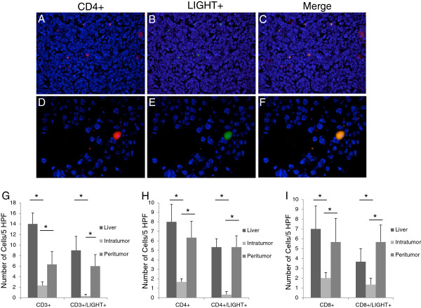
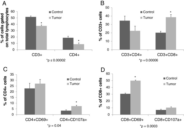
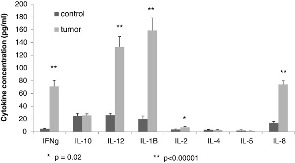
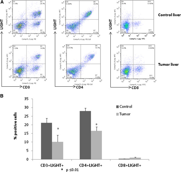
CD3+ and CD4+ lymphocytes were decreased, and CD8+ cells were increased in CRLM compared to control liver. When present, greater populations of tumor infiltrating lymphocytes (TIL) were found peritumoral than intratumoral. The TIL expressed significantly higher levels of CD69 and CD107a, but lower levels of LIGHT. Cytokine expression profiles revealed increased levels of the T-helper 1 (Th1) cytokines IFN gamma, IL-12, IL-1b, and IL-8 in CRLM compared to control liver tissue. There was no difference in T-helper 2 (Th2) cytokines between the groups.
Characterization of the tumor microenvironment of CRLM revealed that although a limited number of activated T-cells infiltrate the tumor and initiate an immune response, the number of LIGHT + T cells infiltrating the tumor were very low. Techniques to decrease suppressive influences or augment the cytotoxic T-cell response are needed and may be possible through mechanisms that can increase intratumoral TIL LIGHT expression.
原发性结肠肿瘤中的 T 细胞浸润与患者生存改善相关。初步数据支持结直肠肝转移瘤(CRLM)中存在类似的关联,并且我们之前发现免疫刺激性细胞因子 LIGHT(TNFSF14)在 CRLM 中的表达增加与患者预后改善相关。因此,增强 CRLM 中 T 细胞反应的机制可能是一种很有前途的治疗方式,然而,CRLM 的肿瘤免疫微环境和 LIGHT 表达仍有待阐明。
利用 CRLM 的同源和免疫相容模型,通过流式细胞术、免疫测定和免疫荧光显微镜,对淋巴细胞表型、功能和位置进行了免疫微环境特征分析。
与对照肝相比,CRLM 中的 CD3+和 CD4+淋巴细胞减少,CD8+细胞增加。当存在时,肿瘤周围的肿瘤浸润淋巴细胞(TIL)比肿瘤内的 TIL 更多。TIL 表达的 CD69 和 CD107a 水平明显更高,而 LIGHT 水平更低。细胞因子表达谱显示,与对照肝组织相比,CRLM 中的 T 辅助 1(Th1)细胞因子 IFNγ、IL-12、IL-1b 和 IL-8 水平升高。两组之间 Th2 细胞因子无差异。
对 CRLM 肿瘤微环境的特征分析表明,尽管有少量活化的 T 细胞浸润肿瘤并引发免疫反应,但浸润肿瘤的 LIGHT+T 细胞数量非常少。需要减少抑制影响或增强细胞毒性 T 细胞反应的技术,并且可能通过能够增加肿瘤内 TIL LIGHT 表达的机制来实现。