Department of Veterinary Pathology, Freie Universität Berlin, Robert-von-Ostertag-Strasse 15, 14163 Berlin, Germany.
BMC Vet Res. 2010 Mar 15;6:15. doi: 10.1186/1746-6148-6-15.
Cell adhesion is an important regulator of cell growth and motility. Recently the hepatocyte cell adhesion molecules 1 and 2 (HEPACAM1 and 2), members of the immunoglobulin family of adhesion genes, have been identified. HEPACAM1 is involved in negative cell cycle regulation via p53, p21 and p27 signalling but also mediates increased human breast cancer cell spread. The role and expression pattern of HEPACAM2 has not been analyzed so far. In the present study we quantified gene expression levels of HEPACAM1 and 2 to evaluate their possible role during the carcinogenesis of canine mammary tumours.
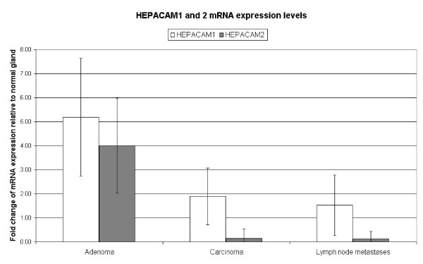
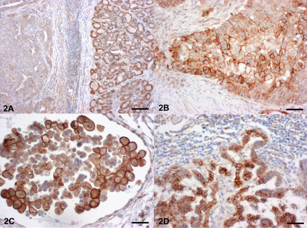
Adenomas displayed increased HEPACAM1 and 2 mRNA expression levels and decreased HEPACAM1 protein expression levels when compared to normal gland, carcinomas and lymph node metastases. In contrast, metastatic carcinomas, intravascular tumour cells and lymph node metastases had HEPACAM 1 protein and mRNA expression levels similar to normal gland but decreased HEPACAM2 mRNA expression when compared to normal gland of the same dog.
HEPACAM1 and 2 seem to be important for cell-cell adhesion of normal and neoplastic canine mammary cells. The loss of HEPACAM1 protein expression in adenomas but not in carcinomas questions its role as a tumour suppressor at late stages of malignant transformation and indicates that it might rather be involved in physiologic mammary cell adhesion and canine mammary tumour metastasis. Furthermore, it can be speculated, whether HEPACAM2 plays a different role in malignancy and metastasis of canine mammary tumours since its transcriptional levels are different in carcinomas and their lymph node metastases when compared to HEPACAM1.
细胞黏附是细胞生长和迁移的重要调节因子。最近,已鉴定出属于免疫球蛋白家族黏附基因的肝细胞黏附分子 1 和 2(HEPACAM1 和 2)。HEPACAM1 通过 p53、p21 和 p27 信号通路参与负性细胞周期调控,但也介导人乳腺癌细胞侵袭性增加。HEPACAM2 的作用和表达模式尚未得到分析。在本研究中,我们定量分析了 HEPACAM1 和 2 的基因表达水平,以评估它们在犬乳腺肿瘤发生过程中的可能作用。
与正常组织相比,腺瘤显示 HEPACAM1 和 2 mRNA 表达水平升高,HEPACAM1 蛋白表达水平降低,而转移性癌、血管内肿瘤细胞和淋巴结转移与正常组织相比具有相似的 HEPACAM1 蛋白和 mRNA 表达水平,但与同一只犬的正常组织相比,HEPACAM2 mRNA 表达水平降低。
HEPACAM1 和 2 似乎对正常和肿瘤犬乳腺细胞的细胞间黏附很重要。HEPACAM1 蛋白在腺瘤中的表达缺失,但在癌组织中不缺失,这使其作为肿瘤抑制因子在恶性转化的晚期阶段的作用受到质疑,并表明其可能更多地参与生理乳腺细胞黏附和犬乳腺肿瘤转移。此外,由于其转录水平在癌组织及其淋巴结转移中与 HEPACAM1 不同,因此可以推测 HEPACAM2 在犬乳腺肿瘤的恶性程度和转移中发挥不同的作用。