ection of Endocrinology and Diabetes, University of Oklahoma Health Sciences Center, Oklahoma City, Oklahoma, USA.
Diabetes. 2010 Jun;59(6):1386-96. doi: 10.2337/db09-1637. Epub 2010 Mar 18.
The oxidation of LDLs is considered a key step in the development of atherosclerosis. How LDL oxidation contributes to atherosclerosis remains poorly defined. Here we report that oxidized and glycated LDL (HOG-LDL) causes aberrant endoplasmic reticulum (ER) stress and that the AMP-activated protein kinase (AMPK) suppressed HOG-LDL-triggered ER stress in vivo.
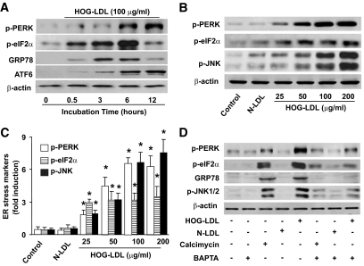
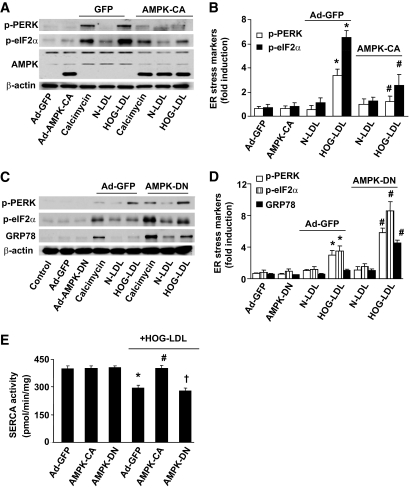
ER stress markers, sarcoplasmic/endoplasmic reticulum Ca(2+) ATPase (SERCA) activity and oxidation, and AMPK activity were monitored in cultured bovine aortic endothelial cells (BAECs) exposed to HOG-LDL or in isolated aortae from mice fed an atherogenic diet.
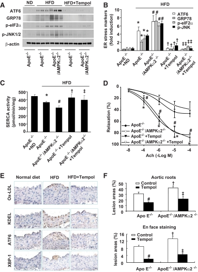
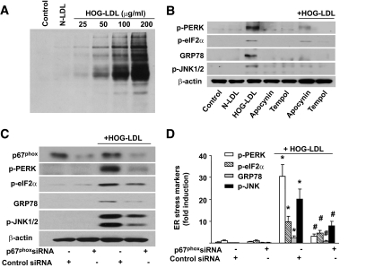
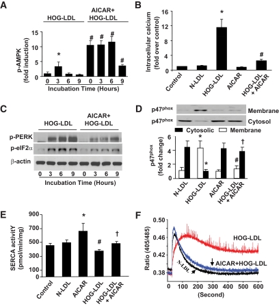
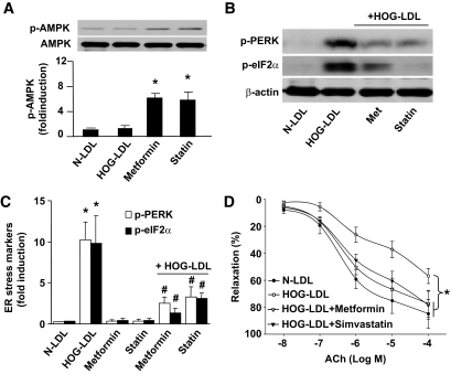
Exposure of BAECs to clinically relevant concentrations of HOG-LDL induced prolonged ER stress and reduced SERCA activity but increased SERCA oxidation. Chronic administration of Tempol (a potent antioxidant) attenuated both SERCA oxidation and aberrant ER stress in mice fed a high-fat diet in vivo. Likewise, AMPK activation by pharmacological (5'-aminoimidazole-4-carboxymide-1-beta-d-ribofuranoside, metformin, and statin) or genetic means (adenoviral overexpression of constitutively active AMPK mutants) significantly mitigated ER stress and SERCA oxidation and improved the endothelium-dependent relaxation in isolated mouse aortae. Finally, Tempol administration markedly attenuated impaired endothelium-dependent vasorelaxation, SERCA oxidation, ER stress, and atherosclerosis in ApoE(-/-) and ApoE(-/-)/AMPKalpha2(-/-) fed a high-fat diet.
We conclude that HOG-LDL, via enhanced SERCA oxidation, causes aberrant ER stress, endothelial dysfunction, and atherosclerosis in vivo, all of which are inhibited by AMPK activation.
LDL 的氧化被认为是动脉粥样硬化发展的关键步骤。LDL 氧化如何导致动脉粥样硬化仍不清楚。在这里,我们报告氧化和糖化的 LDL(HOG-LDL)引起内质网(ER)应激异常,并且 AMP 激活蛋白激酶(AMPK)在体内抑制 HOG-LDL 触发的 ER 应激。
在暴露于 HOG-LDL 的培养牛主动脉内皮细胞(BAEC)或在喂食动脉粥样硬化饮食的小鼠分离的主动脉中监测 ER 应激标志物、肌浆/内质网 Ca(2+)ATP 酶(SERCA)活性和氧化以及 AMPK 活性。
暴露于临床相关浓度的 HOG-LDL 的 BAEC 诱导延长的 ER 应激和降低的 SERCA 活性,但增加 SERCA 氧化。体内给予 Tempo(一种有效的抗氧化剂)在喂食高脂肪饮食的小鼠中减弱了 SERCA 氧化和异常的 ER 应激。同样,通过药理学(5'-氨基咪唑-4-羧基酰胺-1-β-d-核糖呋喃糖苷、二甲双胍和他汀类药物)或遗传手段(腺病毒过表达组成型激活的 AMPK 突变体)激活 AMPK 可显著减轻 ER 应激和 SERCA 氧化,并改善分离的小鼠主动脉中的内皮依赖性松弛。最后,Tempo 给药显著减弱了高脂饮食喂养的 ApoE(-/-)和 ApoE(-/-)/AMPKalpha2(-/-)小鼠中受损的内皮依赖性血管舒张、SERCA 氧化、ER 应激和动脉粥样硬化。
我们得出结论,HOG-LDL 通过增强 SERCA 氧化,在体内引起内质网应激异常、内皮功能障碍和动脉粥样硬化,所有这些都被 AMPK 激活所抑制。