• 文献检索
  • 文档翻译
  • 深度研究
  • 学术资讯
  • Suppr Zotero 插件Zotero 插件
  • 邀请有礼
  • 套餐&价格
  • 历史记录
应用&插件
Suppr Zotero 插件Zotero 插件浏览器插件Mac 客户端Windows 客户端微信小程序
定价
高级版会员购买积分包购买API积分包
服务
文献检索文档翻译深度研究API 文档MCP 服务
关于我们
关于 Suppr公司介绍联系我们用户协议隐私条款
关注我们

Suppr 超能文献

核心技术专利:CN118964589B侵权必究
粤ICP备2023148730 号-1Suppr @ 2026

文献检索

告别复杂PubMed语法,用中文像聊天一样搜索,搜遍4000万医学文献。AI智能推荐,让科研检索更轻松。

立即免费搜索

文件翻译

保留排版,准确专业,支持PDF/Word/PPT等文件格式,支持 12+语言互译。

免费翻译文档

深度研究

AI帮你快速写综述,25分钟生成高质量综述,智能提取关键信息,辅助科研写作。

立即免费体验

用于分辨交叉纤维的最佳 b 值:使用 1.5-T MR 进行标准临床 b 值研究。

Optimum b value for resolving crossing fibers: a study with standard clinical b value using 1.5-T MR.

机构信息

Department of Radiology, Graduate School of Medical Science, Kyoto Prefectural University of Medicine, Kajii-cho, Kawaramachi Hirokoji Agaru, Kamigyo-ku, Kyoto City, Kyoto, 602-8566, Japan.

出版信息

Neuroradiology. 2010 Aug;52(8):723-8. doi: 10.1007/s00234-010-0670-0. Epub 2010 Mar 23.

DOI:10.1007/s00234-010-0670-0
PMID:20309533
原文链接:https://pmc.ncbi.nlm.nih.gov/articles/PMC2901494/
Abstract

INTRODUCTION

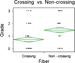
We sought to investigate the optimum b value for resolving crossing fiber using high-angular resolution diffusion imaging (HARDI)-based multi-tensor tractography. The study tested the standard b values that are commonly used in the routine clinical setting.

METHODS

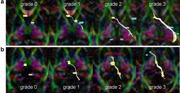
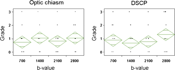
Ten normal volunteers (five men and five women) with a mean age of 26.3 years (range, 22-32 years) were scanned using a 1.5-T clinical magnetic resonance unit. Single-shot echo-planar imaging was used for diffusion-weighted imaging with a diffusion-sensitizing gradient in 32 orientations. The b values of 700, 1,400, 2,100, and 2,800 s/m(2) were used. Data postprocessing was performed using multi-tensor methods. The depiction of the optic nerves, optic tracts, and decussation of superior cerebellar peduncles were assessed.

RESULTS

The depictions of the nerve fibers were independent of the b values tested.

CONCLUSION

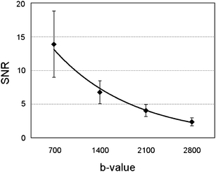
The depiction of crossing fibers by HARDI-based multi-tensor tractography is not substantially influenced by b values ranging from 700 to 2,800 s/m(2). Thus, the optimum b value within this range may be the lowest one considering the higher signal to noise ratio.

摘要

简介

我们旨在研究使用基于高角分辨率扩散成像(HARDI)的多张量追踪技术来解决交叉纤维的最佳 b 值。本研究测试了常规临床设置中常用的标准 b 值。

方法

10 名正常志愿者(男 5 名,女 5 名),平均年龄 26.3 岁(范围 22-32 岁),在 1.5T 临床磁共振仪上进行扫描。单次激发回波平面成像用于扩散加权成像,扩散敏感梯度在 32 个方向上。使用了 700、1400、2100 和 2800 s/m 2 的 b 值。使用多张量方法进行数据后处理。评估了视神经、视束和上小脑脚交叉的描绘。

结果

纤维的描绘与测试的 b 值无关。

结论

基于 HARDI 的多张量追踪技术对交叉纤维的描绘不受 700 至 2800 s/m 2 范围内 b 值的显著影响。因此,考虑到更高的信噪比,该范围内的最佳 b 值可能是最低值。

https://cdn.ncbi.nlm.nih.gov/pmc/blobs/4cef/2901494/3dadfaec28cf/234_2010_670_Fig4_HTML.jpg
https://cdn.ncbi.nlm.nih.gov/pmc/blobs/4cef/2901494/4d5804dc022b/234_2010_670_Fig1_HTML.jpg
https://cdn.ncbi.nlm.nih.gov/pmc/blobs/4cef/2901494/5397427f7f55/234_2010_670_Fig2_HTML.jpg
https://cdn.ncbi.nlm.nih.gov/pmc/blobs/4cef/2901494/d482529c09b4/234_2010_670_Fig3_HTML.jpg
https://cdn.ncbi.nlm.nih.gov/pmc/blobs/4cef/2901494/3dadfaec28cf/234_2010_670_Fig4_HTML.jpg

相似文献

1
Optimum b value for resolving crossing fibers: a study with standard clinical b value using 1.5-T MR.用于分辨交叉纤维的最佳 b 值:使用 1.5-T MR 进行标准临床 b 值研究。
Neuroradiology. 2010 Aug;52(8):723-8. doi: 10.1007/s00234-010-0670-0. Epub 2010 Mar 23.
2
Diffusion tensor fiber tractography of the optic radiation: analysis with 6-, 12-, 40-, and 81-directional motion-probing gradients, a preliminary study.视辐射的扩散张量纤维束成像:使用6、12、40和81方向运动探测梯度的分析,一项初步研究。
AJNR Am J Neuroradiol. 2007 Jan;28(1):92-6.
3
Multitensor tractography enables better depiction of motor pathways: initial clinical experience using diffusion-weighted MR imaging with standard b-value.多张量纤维束成像能够更好地描绘运动通路:使用具有标准b值的扩散加权磁共振成像的初步临床经验。
AJNR Am J Neuroradiol. 2007 Oct;28(9):1668-73. doi: 10.3174/ajnr.A0640. Epub 2007 Sep 20.
4
Diffusion tensor imaging and fiber tractography of the median nerve at 1.5T: optimization of b value.1.5T下正中神经的扩散张量成像和纤维束成像:b值的优化
Skeletal Radiol. 2009 Jan;38(1):51-9. doi: 10.1007/s00256-008-0577-6. Epub 2008 Sep 5.
5
Estimation of the anterior extent of the Meyer loop using MR tractography.使用磁共振神经成像术估计迈耶袢的前部范围。
AJNR Am J Neuroradiol. 2008 Mar;29(3):E4; auhtor reply E5. doi: 10.3174/ajnr.A0889. Epub 2008 Jan 2.
6
Resolving fiber crossing using advanced fast marching tractography based on diffusion tensor imaging.基于扩散张量成像的先进快速行进纤维束成像技术解决纤维交叉问题。
Neuroimage. 2006 Mar;30(1):110-20. doi: 10.1016/j.neuroimage.2005.09.027. Epub 2005 Oct 24.
7
High-resolution diffusion tensor imaging and tractography of the human optic chiasm at 9.4 T.9.4T下人类视交叉的高分辨率扩散张量成像及纤维束成像
Neuroimage. 2008 Jan 1;39(1):157-68. doi: 10.1016/j.neuroimage.2007.08.015. Epub 2007 Aug 24.
8
Diffusion spectrum magnetic resonance imaging (DSI) tractography of crossing fibers.交叉纤维的扩散谱磁共振成像(DSI)纤维束成像
Neuroimage. 2008 Jul 15;41(4):1267-77. doi: 10.1016/j.neuroimage.2008.03.036. Epub 2008 Apr 8.
9
[Optic radiation in normal adults: a study using magnetic resonance diffusion tensor imaging and diffusion tensor tractography].[正常成年人的视辐射:一项使用磁共振扩散张量成像和扩散张量纤维束成像的研究]
Nan Fang Yi Ke Da Xue Xue Bao. 2008 Mar;28(3):396-8.
10
Voxel-based analysis of the diffusion tensor.体素基分析弥散张量。
Neuroradiology. 2010 Aug;52(8):699-710. doi: 10.1007/s00234-010-0716-3. Epub 2010 May 14.

引用本文的文献

1
Nerve fibre organisation in the human optic nerve and chiasm: what do we really know?人类视神经和视交叉中的神经纤维组织:我们到底知道多少?
Eye (Lond). 2024 Aug;38(12):2457-2471. doi: 10.1038/s41433-024-03137-7. Epub 2024 Jun 7.
2
Methods of diffusion MRI tractography for localization of the anterior optic pathway: A systematic review of validated methods.扩散磁共振成像轨迹法在前视路定位中的应用:验证方法的系统评价。
Neuroimage Clin. 2023;39:103494. doi: 10.1016/j.nicl.2023.103494. Epub 2023 Aug 10.
3
Resolution and b value dependent structural connectome in ex vivo mouse brain.

本文引用的文献

1
Probabilistic diffusion tractography with multiple fibre orientations: What can we gain?具有多种纤维取向的概率性扩散张量成像:我们能获得什么?
Neuroimage. 2007 Jan 1;34(1):144-55. doi: 10.1016/j.neuroimage.2006.09.018. Epub 2006 Oct 27.
2
Diffusion tensor imaging and fiber tractography in acute stroke.急性卒中的扩散张量成像和纤维束成像
Neuroimaging Clin N Am. 2005 Aug;15(3):655-65, xii. doi: 10.1016/j.nic.2005.08.010.
3
Resolving fiber crossing using advanced fast marching tractography based on diffusion tensor imaging.
离体小鼠脑的分辨率和 b 值相关结构连接组学
Neuroimage. 2022 Jul 15;255:119199. doi: 10.1016/j.neuroimage.2022.119199. Epub 2022 Apr 10.
4
Diffusion tensor tractography of normal facial and vestibulocochlear nerves.正常面神经和前庭蜗神经的扩散张量纤维束成像
Int J Comput Assist Radiol Surg. 2015 Apr;10(4):383-92. doi: 10.1007/s11548-014-1129-2. Epub 2014 Nov 20.
5
Reconstruction of scattered data in fetal diffusion MRI.胎儿扩散磁共振成像中离散数据的重建。
Med Image Anal. 2012 Jan;16(1):28-37. doi: 10.1016/j.media.2011.04.004. Epub 2011 Apr 27.
6
Dentatorubrothalamic tract in human brain: diffusion tensor tractography study.人脑齿状核红核被盖束:弥散张量纤维束成像研究。
Neuroradiology. 2011 Oct;53(10):787-91. doi: 10.1007/s00234-011-0878-7. Epub 2011 May 3.
基于扩散张量成像的先进快速行进纤维束成像技术解决纤维交叉问题。
Neuroimage. 2006 Mar;30(1):110-20. doi: 10.1016/j.neuroimage.2005.09.027. Epub 2005 Oct 24.
4
Mapping complex tissue architecture with diffusion spectrum magnetic resonance imaging.利用扩散谱磁共振成像绘制复杂组织结构图。
Magn Reson Med. 2005 Dec;54(6):1377-86. doi: 10.1002/mrm.20642.
5
Multitensor approach for analysis and tracking of complex fiber configurations.用于分析和追踪复杂纤维结构的多张量方法
Magn Reson Med. 2005 Nov;54(5):1216-25. doi: 10.1002/mrm.20670.
6
Q-ball imaging.Q球成像
Magn Reson Med. 2004 Dec;52(6):1358-72. doi: 10.1002/mrm.20279.
7
Direct estimation of the fiber orientation density function from diffusion-weighted MRI data using spherical deconvolution.使用球面反卷积从扩散加权磁共振成像数据直接估计纤维取向密度函数。
Neuroimage. 2004 Nov;23(3):1176-85. doi: 10.1016/j.neuroimage.2004.07.037.
8
Quantitative evaluation of image-based distortion correction in diffusion tensor imaging.扩散张量成像中基于图像的失真校正的定量评估。
IEEE Trans Med Imaging. 2004 Jul;23(7):789-98. doi: 10.1109/TMI.2004.827479.
9
High angular resolution diffusion imaging reveals intravoxel white matter fiber heterogeneity.高角分辨率扩散成像揭示了体素内白质纤维的异质性。
Magn Reson Med. 2002 Oct;48(4):577-82. doi: 10.1002/mrm.10268.
10
Diffusion-weighted imaging of cerebral infarctions: are higher B values better?脑梗死的扩散加权成像:更高的B值会更好吗?
J Comput Assist Tomogr. 2002 Jul-Aug;26(4):622-7. doi: 10.1097/00004728-200207000-00026.