Department of Orthopaedic Surgery, University Hospital Carl Gustav Carus, Medical Faculty of the Technical University of Dresden, Dresden, Germany.
BMC Musculoskelet Disord. 2010 Mar 25;11:57. doi: 10.1186/1471-2474-11-57.
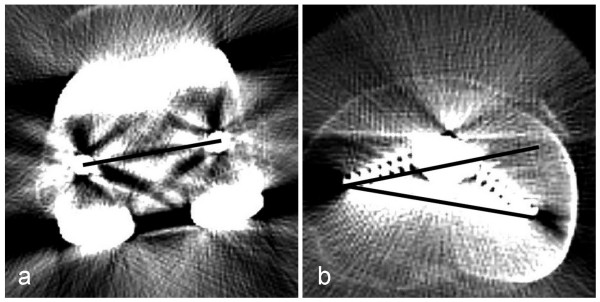
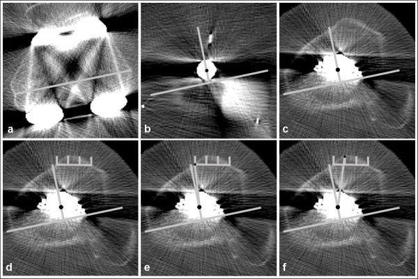
Correct rotational alignment of the femoral and tibial component is an important factor for successful TKA. The transepicondylar axis is widely accepted as a reference for the femoral component. There is not a standard reference for the tibial component. CT scans were used in this study to measure which of 2 tibial landmarks most reliably reproduces a correct femoro-tibial rotational alignment in TKA.
80 patients received a cemented, unconstrained, cruciate-retaining TKA with a rotating platform. CT scans were performed 5-7 days postoperatively but before discharge. The rotational mismatch between the femoral and tibial components was measured. Furthermore, the rotational variance between the transepicondylar line, as a reference for the orientation of the femoral component and different tibial landmarks, was measured.
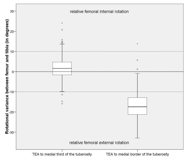
There was notable rotational mismatch between the femoral and tibial components. The median mismatch was 0 degrees (range: 16.2 degrees relative external to 14.4 degrees relative internal rotation of the femoral component).Using the transepicondylar line as a reference for femoral rotational alignment and the medial third of the tuberosity as a reference for tibial rotational alignment, 67.5% of all TKA had a femoro-tibial variance within +/- 5 degrees, 85% within +/- 10 degrees and 97.5% within +/- 20 degrees. Using the medial border of the tibial tubercle as a reference this variance was greater, only 3.8% had a femoro-tibial variance within +/- 5 degrees, 15% within +/- 10 degrees and 68.8% within +/- 20 degrees.
Using fixed bone landmarks for rotational alignment leads to a notable variance between femoral and tibial components. Referencing the tibial rotation on a line from the medial third of the tibial tubercle to the center of the tibial tray resulted in a better femoro-tibial rotational alignment than using the medial border of tibial tubercle as a landmark. Surgeons using fixed bearings with a high rotational constraint between the inlay and the femoral component should be aware of this effect to avoid premature polyethylene wear.
Clinical trials registry NCT01022099.
股骨和胫骨组件的正确旋转对线是全膝关节置换术(TKA)成功的一个重要因素。髁间轴被广泛认为是股骨组件的参考。但是,胫骨组件没有标准的参考。本研究使用 CT 扫描来测量在 TKA 中,2 个胫骨标志中哪一个最能可靠地再现正确的股骨-胫骨旋转对线。
80 例患者接受了骨水泥固定、非约束性、保留十字韧带的旋转平台 TKA。术后 5-7 天(即在出院前)进行 CT 扫描。测量股骨和胫骨组件之间的旋转不匹配。此外,还测量了髁间线(作为股骨组件方向的参考)和不同胫骨标志之间的旋转差异。
股骨和胫骨组件之间存在明显的旋转不匹配。中位数不匹配为 0 度(股骨组件相对外旋 16.2 度,相对内旋 14.4 度)。使用髁间线作为股骨旋转对线的参考,使用胫骨结节的内三分之一作为胫骨旋转对线的参考,所有 TKA 中有 67.5%的股骨-胫骨差异在 +/- 5 度以内,85%在 +/- 10 度以内,97.5%在 +/- 20 度以内。使用胫骨结节的内侧缘作为参考,这种差异更大,只有 3.8%的股骨-胫骨差异在 +/- 5 度以内,15%在 +/- 10 度以内,68.8%在 +/- 20 度以内。
使用固定的骨标志进行旋转对线会导致股骨和胫骨组件之间存在明显的差异。将胫骨旋转参考线从胫骨结节的内三分之一到胫骨托的中心,与使用胫骨结节的内侧缘作为标志相比,可获得更好的股骨-胫骨旋转对线。使用具有高旋转约束的固定衬垫的外科医生应该意识到这种影响,以避免过早的聚乙烯磨损。
临床试验注册 NCT01022099。