Electron Microscopy (EM) Facility, National Institute of Neurological Disorders and Stroke (NINDS), National Institutes of Health (NIH), Bethesda, MD, USA.
Neuroscience. 2010 Jun 16;168(1):11-7. doi: 10.1016/j.neuroscience.2010.03.041. Epub 2010 Mar 25.
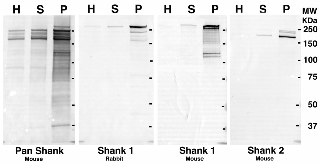
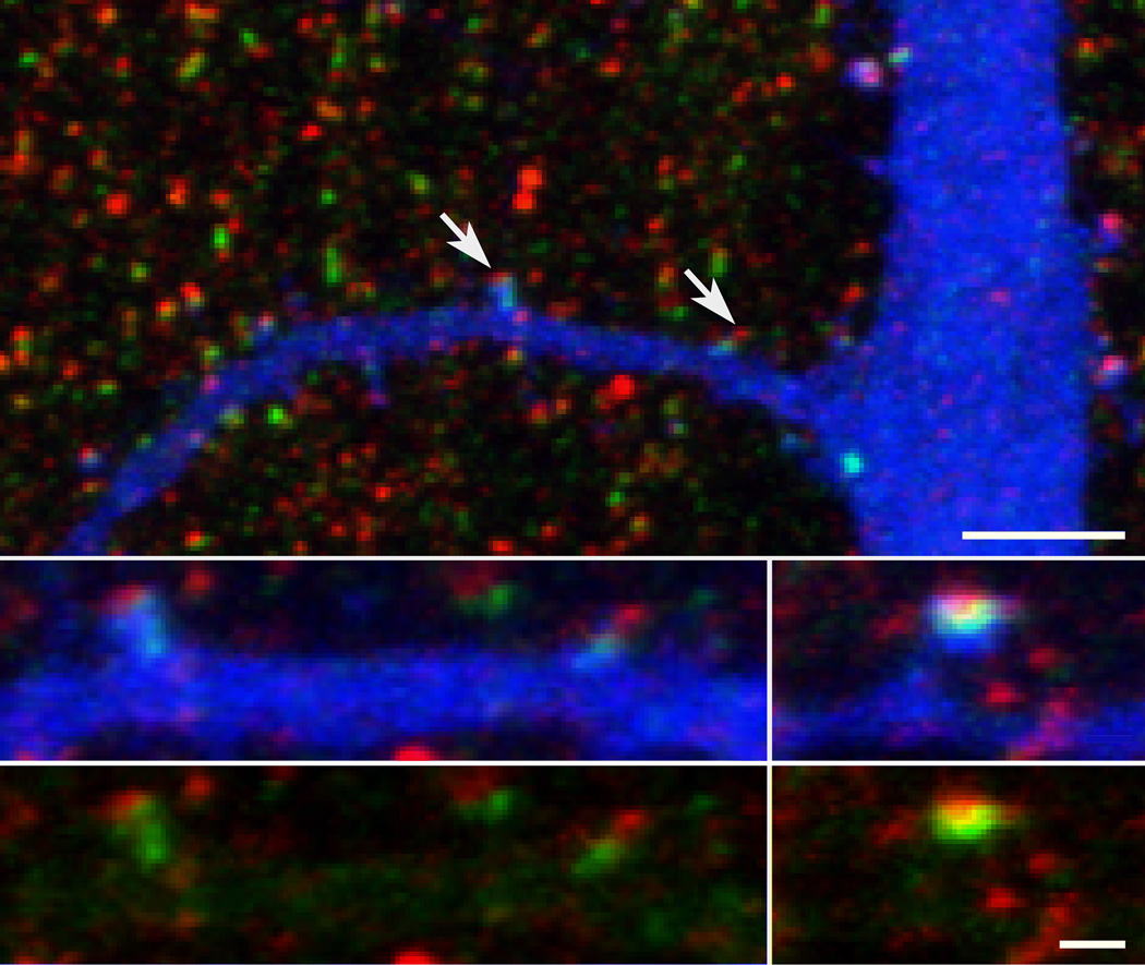
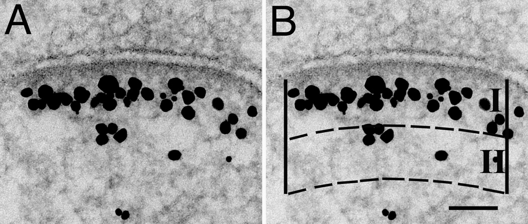
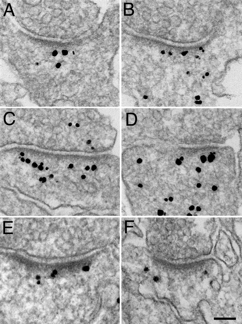
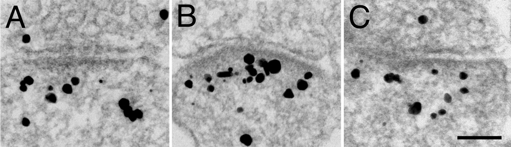
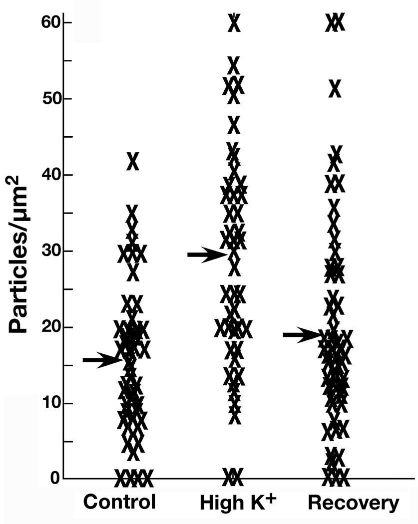
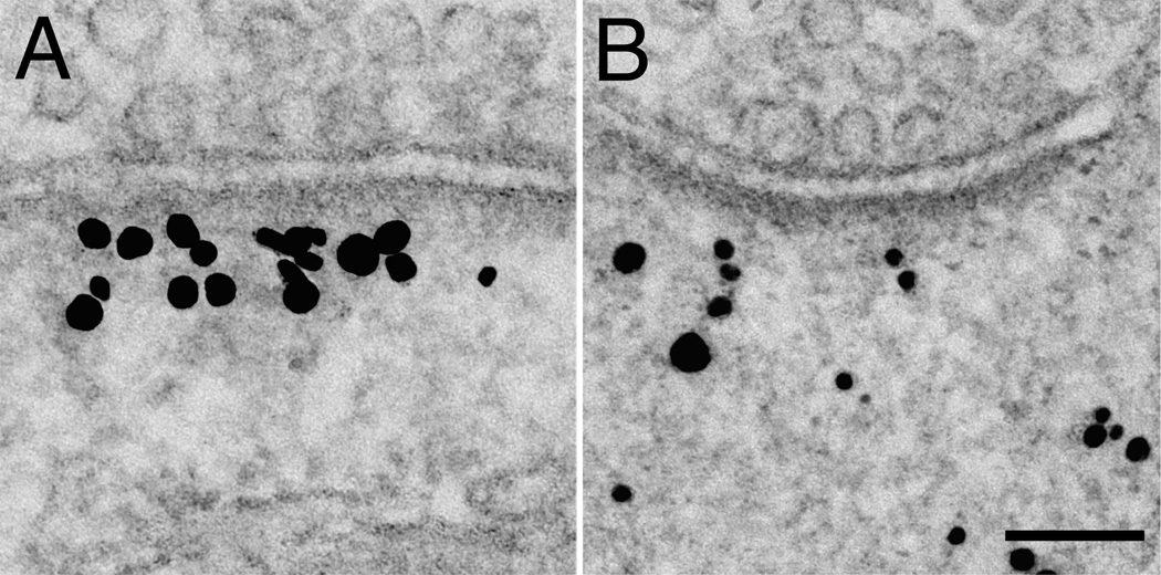
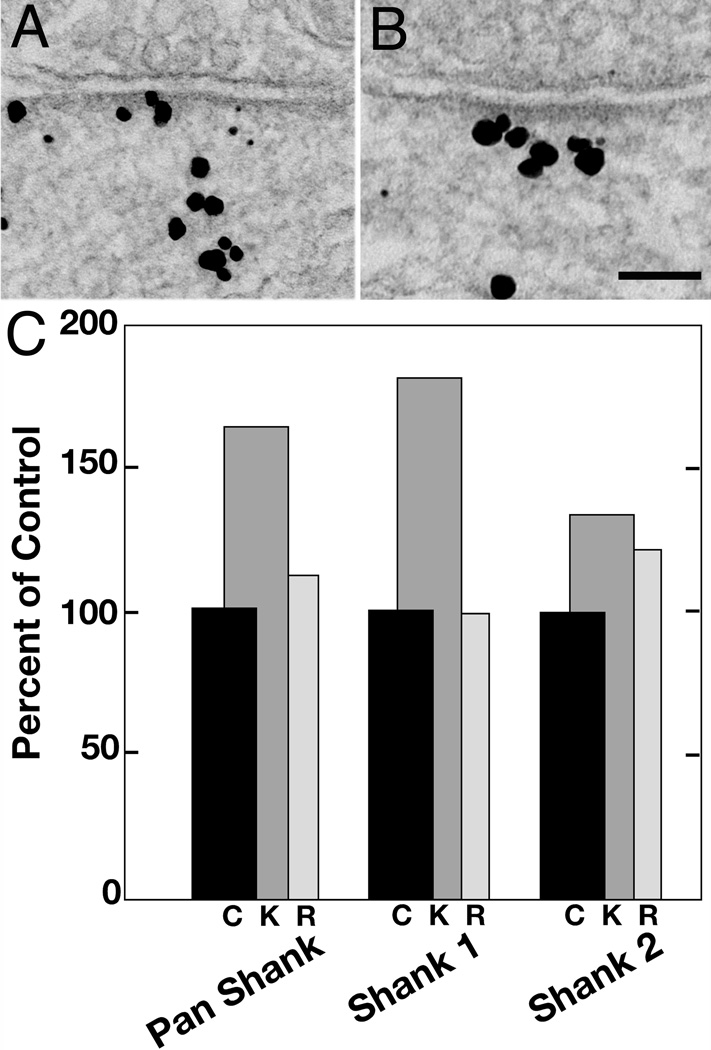
Dendritic spines contain a family of abundant scaffolding proteins known as Shanks, but little is known about how their distributions might change during synaptic activity. Here, pre-embedding immunogold electron microscopy is used to localize Shanks in synapses from cultured hippocampal neurons. We find that Shanks are preferentially located at postsynaptic densities (PSDs) as well as in a filamentous network near the PSD, extending up to 120 nm from the postsynaptic membrane. Application of sub-type specific antibodies shows that Shank2 is typically concentrated at and near PSDs while Shank1 is, in addition, distributed throughout the spine head. Depolarization with high K+ for 2 min causes transient, reversible translocation of Shanks towards the PSD that is dependent on extracellular Ca2+. The amount of activity-induced redistribution and subsequent recovery is pronounced for Shank1 but less so for Shank2. Thus, Shank1 appears to be a dynamic element within the spine, whose translocation could be involved in activity-induced, transient structural changes, while Shank2 appears to be a more stable element positioned at the interface of the PSD with the spine cytoplasm.
树突棘含有大量支架蛋白家族,称为 Shank,但对于它们在突触活动期间的分布如何变化知之甚少。在这里,使用预嵌入免疫胶体金电子显微镜技术在培养的海马神经元突触中定位 Shank。我们发现 Shank 优先位于突触后密度(PSD)以及 PSD 附近的丝状网络中,从突触后膜延伸至 120nm。应用亚型特异性抗体表明,Shank2 通常集中在 PSD 附近,而 Shank1 除了分布在整个棘突头部之外。用高 K+ 去极化 2 分钟会导致 Shanks 向 PSD 的瞬时、可逆易位,这依赖于细胞外 Ca2+。Shank1 的活性诱导重分布的量和随后的恢复量较大,但 Shank2 的则较小。因此,Shank1 似乎是棘突内的一个动态元件,其易位可能与活性诱导的短暂结构变化有关,而 Shank2 似乎是位于 PSD 与棘突细胞质界面的更稳定元件。