Tao-Cheng Jung-Hwa, Toy Dana, Winters Christine A, Reese Thomas S, Dosemeci Ayse
NINDS EM Facility, National Institute of Neurological Disorders and Stroke, National Institutes of Health, Bethesda, MD, United States of America.
Laboratory of Neurobiology, National Institute of Neurological Disorders and Stroke, National Institutes of Health, Bethesda, MD, United States of America.
PLoS One. 2016 May 4;11(5):e0153979. doi: 10.1371/journal.pone.0153979. eCollection 2016.
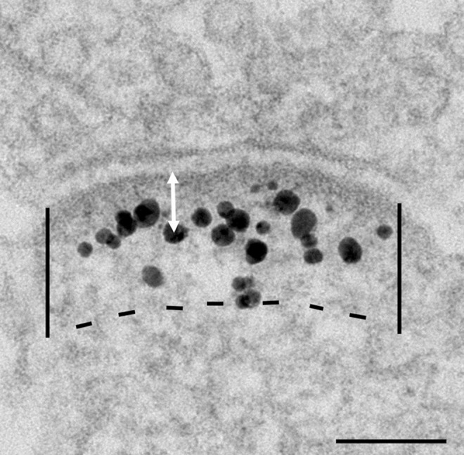
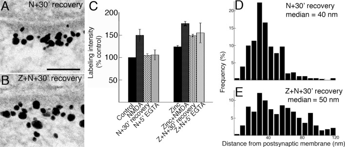
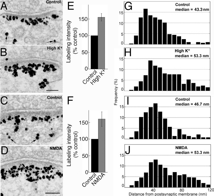
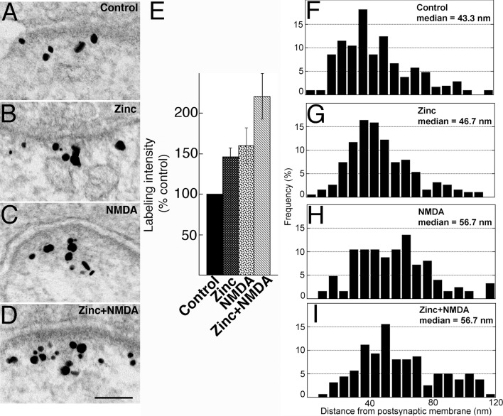
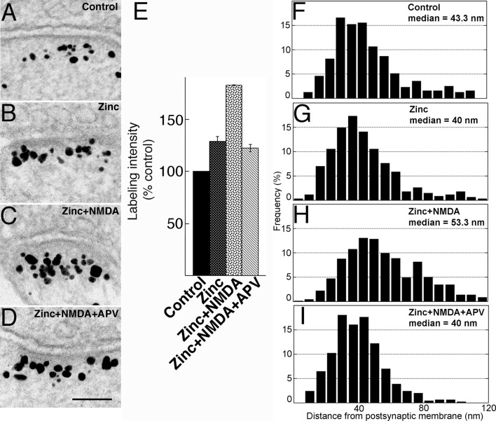
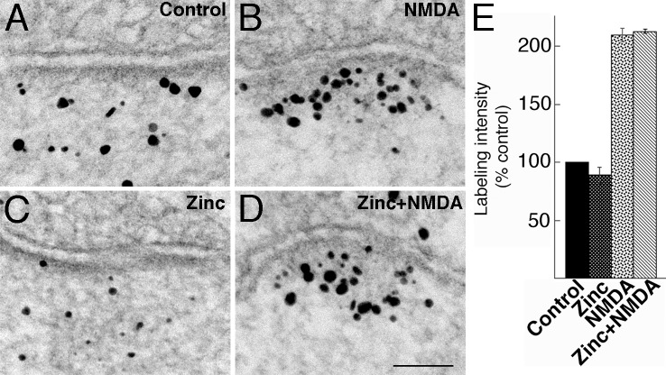
Shank3 is a postsynaptic density (PSD) scaffold protein of the Shank family. Here we use pre-embedding immunogold electron microscopy to investigate factors influencing the distribution of Shank3 at the PSD. In dissociated rat hippocampal cultures under basal conditions, label for Shank3 was concentrated in a broad layer of the PSD, ~20-80 nm from the postsynaptic membrane. Upon depolarization with high K+ (90 mM, 2 min), or application of NMDA (50 μM, 2 min), both the labeling intensity at the PSD and the median distance of label from the postsynaptic membrane increased significantly, indicating that Shank3 molecules are preferentially recruited to the distal layer of the PSD. Incubation in medium supplemented with zinc (50 μM ZnCl2, 1 hr) also significantly increased labeling intensity for Shank3 at the PSD, but this addition of Shank3 was not preferential to the distal layer. When cells were incubated with zinc and then treated with NMDA, labeling intensity of Shank3 became higher than with either treatment alone and manifested a preference for the distal layer of the PSD. Without zinc supplementation, NMDA-induced accumulation of Shank3 at the PSD was transient, reversing within 30 min after return to control medium. However, when zinc was included in culture media throughout the experiment, the NMDA-induced accumulation of Shank3 was largely retained, including Shank3 molecules recruited to the distal layer of the PSD. These results demonstrate that activity induces accumulation of Shank3 at the PSD and that zinc stabilizes PSD-associated Shank3, possibly through strengthening of Shank-Shank association.
Shank3是Shank家族的一种突触后致密区(PSD)支架蛋白。在此,我们使用包埋前免疫金电子显微镜来研究影响Shank3在PSD分布的因素。在基础条件下的离体大鼠海马培养物中,Shank3的标记集中在PSD的一个较宽层中,距离突触后膜约20 - 80纳米。在用高钾(90 mM,2分钟)去极化或应用NMDA(50 μM,2分钟)后,PSD处的标记强度以及标记距突触后膜的中位距离均显著增加,这表明Shank3分子优先被招募到PSD的远端层。在补充锌(50 μM ZnCl2,1小时)的培养基中孵育也显著增加了PSD处Shank3的标记强度,但这种Shank3的添加并非优先作用于远端层。当细胞用锌孵育然后用NMDA处理时,Shank3的标记强度高于单独进行任何一种处理,并且表现出对PSD远端层的偏好。在不补充锌的情况下,NMDA诱导的Shank3在PSD处的积累是短暂的,在回到对照培养基后30分钟内就会逆转。然而,当在整个实验过程中培养基中都含有锌时,NMDA诱导的Shank3积累在很大程度上得以保留,包括被招募到PSD远端层的Shank3分子。这些结果表明,活性诱导Shank3在PSD处积累,并且锌稳定与PSD相关的Shank3,可能是通过加强Shank - Shank相互作用来实现的。