Department of Cell Biology & Anatomy, Hotchkiss Brain Institute, University of Calgary, Calgary, Alberta, Canada T2N 4N1.
Cerebellum. 2010 Sep;9(3):352-74. doi: 10.1007/s12311-010-0168-7.
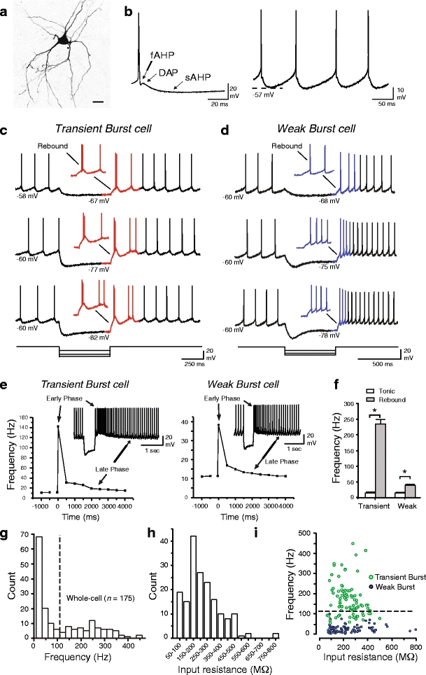
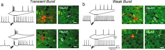
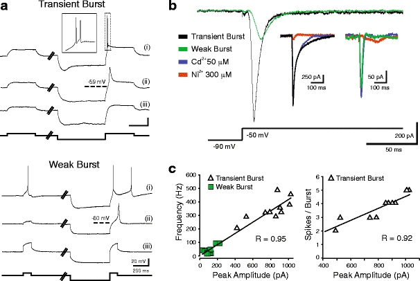
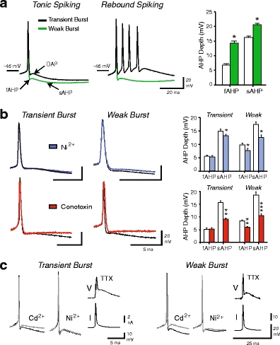
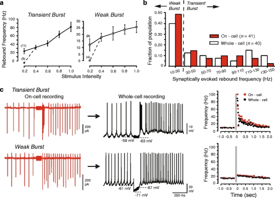
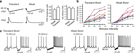
Neurons of the deep cerebellar nuclei (DCN) play a critical role in defining the output of cerebellum in the course of encoding Purkinje cell inhibitory inputs. The earliest work performed with in vitro preparations established that DCN cells have the capacity to translate membrane hyperpolarizations into a rebound increase in firing frequency. The primary means of distinguishing between DCN neurons has been according to cell size and transmitter phenotype, but in some cases, differences in the firing properties of DCN cells maintained in vitro have been reported. In particular, it was shown that large diameter cells in the rat DCN exhibit two phenotypes of rebound discharge in vitro that may eventually help define their functional roles in cerebellar output. A transient burst and weak burst phenotype can be distinguished based on the frequency and pattern of rebound discharge immediately following a hyperpolarizing stimulus. Work to date indicates that the difference in excitability arises from at least the degree of activation of T-type Ca(2+) current during the immediate phase of rebound firing and Ca(2+)-dependent K(+) channels that underlie afterhyperpolarizations. Both phenotypes can be detected following stimulation of Purkinje cell inhibitory inputs under conditions that preserve resting membrane potential and natural ionic gradients. In this paper, we review the evidence supporting the existence of different rebound phenotypes in DCN cells and the ion channel expression patterns that underlie their generation.
小脑深部核(DCN)神经元在编码浦肯野细胞抑制性输入的过程中对小脑的输出起着至关重要的作用。最早使用体外制剂进行的工作表明,DCN 细胞具有将膜超极化转化为放电频率反弹增加的能力。区分 DCN 神经元的主要方法是根据细胞大小和递质表型,但在某些情况下,体外培养的 DCN 细胞的放电特性存在差异。特别是,已经表明大鼠 DCN 中的大直径细胞表现出两种体外反弹放电的表型,这可能最终有助于确定它们在小脑输出中的功能作用。根据超极化刺激后反弹放电的频率和模式,可以区分瞬态爆发和弱爆发表型。迄今为止的工作表明,兴奋性的差异至少来自于反弹放电即刻阶段 T 型 Ca(2+)电流的激活程度以及构成后超极化的 Ca(2+)-依赖性 K(+)通道。在保持静息膜电位和自然离子梯度的条件下,刺激浦肯野细胞抑制性输入后,两种表型都可以检测到。本文综述了支持 DCN 细胞中存在不同反弹表型以及为其产生提供基础的离子通道表达模式的证据。