Deutsches Krebsforschungszentrum, Forschungsschwerpunkt Infektion und Krebs, Abteilung Virale Transformationsmechanismen, Heidelberg, Germany.
Mol Cancer. 2010 Apr 20;9:82. doi: 10.1186/1476-4598-9-82.
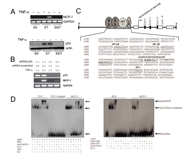
Our previous studies showed that the expression of the monocyte-chemoattractant protein (MCP)-1, a chemokine, which triggers the infiltration and activation of cells of the monocyte-macrophage lineage, is abrogated in human papillomavirus (HPV)-positive premalignant and malignant cells. In silico analysis of the MCP-1 upstream region proposed a putative p53 binding side about 2.5 kb upstream of the transcriptional start. The aim of this study is to monitor a physiological role of p53 in this process.
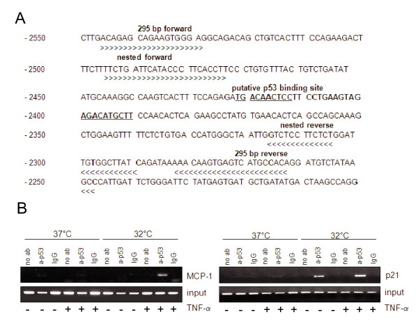
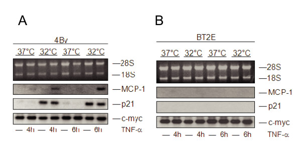
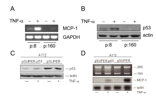
The proposed p53 binding side could be confirmed in vitro by electrophoretic-mobility-shift assays and in vivo by chromatin immunoprecipitation. Moreover, the availability of p53 is apparently important for chemokine regulation, since TNF-alpha can induce MCP-1 only in human keratinocytes expressing the viral oncoprotein E7, but not in HPV16 E6 positive cells, where p53 becomes degraded. A general physiological role of p53 in MCP-1 regulation was further substantiated in HPV-negative cells harboring a temperature-sensitive mutant of p53 and in Li-Fraumeni cells, carrying a germ-line mutation of p53. In both cases, non-functional p53 leads to diminished MCP-1 transcription upon TNF-alpha treatment. In addition, siRNA directed against p53 decreased MCP-1 transcription after TNF-alpha addition, directly confirming a crosstalk between p53 and MCP-1.
These data support the concept that p53 inactivation during carcinogenesis also affects immune surveillance by interfering with chemokine expression and in turn communication with cells of the immunological compartment.
我们之前的研究表明,趋化因子单核细胞趋化蛋白-1(MCP-1)的表达被阻断,该蛋白能触发单核细胞-巨噬细胞系细胞的浸润和激活,在人乳头瘤病毒(HPV)阳性的癌前和恶性细胞中。MCP-1 上游区域的计算机分析提出了一个假定的 p53 结合位点,大约在转录起始点上游 2.5kb 处。本研究的目的是监测 p53 在这个过程中的生理作用。
通过电泳迁移率变动分析可以在体外证实假定的 p53 结合部位,通过染色质免疫沉淀可以在体内证实。此外,p53 的可用性显然对趋化因子的调节很重要,因为 TNF-α 只能诱导 HPV16 E7 表达的人类角质形成细胞中 MCP-1 的产生,而不能诱导 HPV16 E6 阳性细胞中 MCP-1 的产生,在 HPV16 E6 阳性细胞中,p53 被降解。在 HPV 阴性细胞中,p53 携带温度敏感型突变,在携带 p53 种系突变的 Li-Fraumeni 细胞中,进一步证实了 p53 在 MCP-1 调节中的一般生理作用。在这两种情况下,非功能性 p53 导致 TNF-α 处理后 MCP-1 转录减少。此外,针对 p53 的 siRNA 在添加 TNF-α 后降低了 MCP-1 的转录,直接证实了 p53 与 MCP-1 之间的相互作用。
这些数据支持这样的概念,即在致癌过程中 p53 的失活也会通过干扰趋化因子的表达,并反过来与免疫细胞之间的通讯,影响免疫监视。