• 文献检索
  • 文档翻译
  • 深度研究
  • 学术资讯
  • Suppr Zotero 插件Zotero 插件
  • 邀请有礼
  • 套餐&价格
  • 历史记录
应用&插件
Suppr Zotero 插件Zotero 插件浏览器插件Mac 客户端Windows 客户端微信小程序
定价
高级版会员购买积分包购买API积分包
服务
文献检索文档翻译深度研究API 文档MCP 服务
关于我们
关于 Suppr公司介绍联系我们用户协议隐私条款
关注我们

Suppr 超能文献

核心技术专利:CN118964589B侵权必究
粤ICP备2023148730 号-1Suppr @ 2026

文献检索

告别复杂PubMed语法,用中文像聊天一样搜索,搜遍4000万医学文献。AI智能推荐,让科研检索更轻松。

立即免费搜索

文件翻译

保留排版,准确专业,支持PDF/Word/PPT等文件格式,支持 12+语言互译。

免费翻译文档

深度研究

AI帮你快速写综述,25分钟生成高质量综述,智能提取关键信息,辅助科研写作。

立即免费体验

在体外缺血模型中,间充质干细胞通过直接的细胞间连接挽救心肌母细胞免于细胞死亡。

Mesenchymal stem cells rescue cardiomyoblasts from cell death in an in vitro ischemia model via direct cell-to-cell connections.

作者信息

Cselenyák Attila, Pankotai Eszter, Horváth Eszter M, Kiss Levente, Lacza Zsombor

机构信息

Institute of Human Physiology and Clinical Experimental Research, Semmelweis University, H-1094, Tuzoltó utca 37-47, Budapest, Hungary.

出版信息

BMC Cell Biol. 2010 Apr 20;11:29. doi: 10.1186/1471-2121-11-29.

DOI:10.1186/1471-2121-11-29
PMID:20406471
原文链接:https://pmc.ncbi.nlm.nih.gov/articles/PMC2869333/
Abstract

BACKGROUND

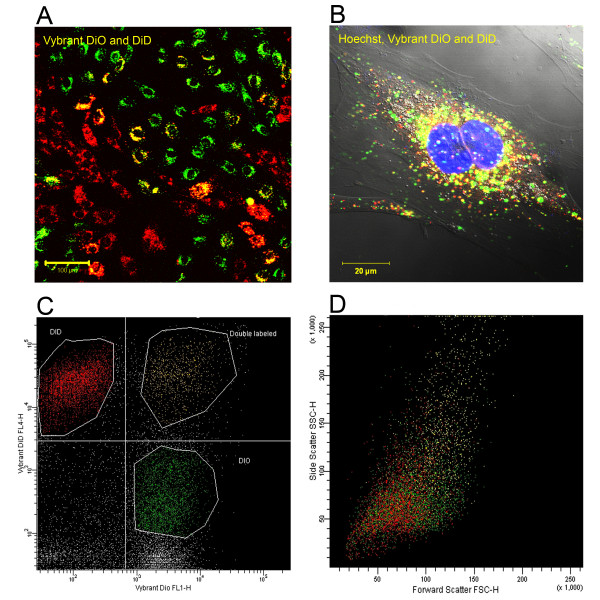
Bone marrow derived mesenchymal stem cells (MSCs) are promising candidates for cell based therapies in myocardial infarction. However, the exact underlying cellular mechanisms are still not fully understood. Our aim was to explore the possible role of direct cell-to-cell interaction between ischemic H9c2 cardiomyoblasts and normal MSCs. Using an in vitro ischemia model of 150 minutes of oxygen glucose deprivation we investigated cell viability and cell interactions with confocal microscopy and flow cytometry.

RESULTS

Our model revealed that adding normal MSCs to the ischemic cell population significantly decreased the ratio of dead H9c2 cells (H9c2 only: 0.85 +/- 0.086 vs. H9c2+MSCs: 0.16 +/- 0.035). This effect was dependent on direct cell-to-cell contact since co-cultivation with MSCs cultured in cell inserts did not exert the same beneficial effect (ratio of dead H9c2 cells: 0.90 +/- 0.055). Confocal microscopy revealed that cardiomyoblasts and MSCs frequently formed 200-500 nm wide intercellular connections and cell fusion rarely occurred between these cells.

CONCLUSION

Based on these results we hypothesize that mesenchymal stem cells may reduce the number of dead cardiomyoblasts after ischemic damage via direct cell-to-cell interactions and intercellular tubular connections may play an important role in these processes.

摘要

背景

骨髓间充质干细胞(MSCs)是心肌梗死细胞治疗中很有前景的候选细胞。然而,确切的潜在细胞机制仍未完全了解。我们的目的是探讨缺血性H9c2心肌母细胞与正常MSCs之间直接细胞间相互作用的可能作用。使用150分钟氧葡萄糖剥夺的体外缺血模型,我们通过共聚焦显微镜和流式细胞术研究了细胞活力和细胞相互作用。

结果

我们的模型显示,向缺血细胞群体中添加正常MSCs可显著降低死亡H9c2细胞的比例(仅H9c2:0.85±0.086 vs. H9c2 + MSCs:0.16±0.035)。这种效应依赖于直接的细胞间接触,因为与培养在细胞插入物中的MSCs共培养没有产生相同的有益效果(死亡H9c2细胞的比例:0.90±0.055)。共聚焦显微镜显示,心肌母细胞和MSCs经常形成200 - 500纳米宽的细胞间连接,并且这些细胞之间很少发生细胞融合。

结论

基于这些结果,我们假设间充质干细胞可能通过直接的细胞间相互作用减少缺血损伤后死亡心肌母细胞的数量,并且细胞间管状连接可能在这些过程中起重要作用。

https://cdn.ncbi.nlm.nih.gov/pmc/blobs/5d97/2869333/1b92416761ae/1471-2121-11-29-6.jpg
https://cdn.ncbi.nlm.nih.gov/pmc/blobs/5d97/2869333/d81ef8dc75fd/1471-2121-11-29-1.jpg
https://cdn.ncbi.nlm.nih.gov/pmc/blobs/5d97/2869333/a969077e730b/1471-2121-11-29-2.jpg
https://cdn.ncbi.nlm.nih.gov/pmc/blobs/5d97/2869333/39fc4561141b/1471-2121-11-29-3.jpg
https://cdn.ncbi.nlm.nih.gov/pmc/blobs/5d97/2869333/3c9aabe57feb/1471-2121-11-29-4.jpg
https://cdn.ncbi.nlm.nih.gov/pmc/blobs/5d97/2869333/84d08249ae2c/1471-2121-11-29-5.jpg
https://cdn.ncbi.nlm.nih.gov/pmc/blobs/5d97/2869333/1b92416761ae/1471-2121-11-29-6.jpg

相似文献

1
Mesenchymal stem cells rescue cardiomyoblasts from cell death in an in vitro ischemia model via direct cell-to-cell connections.在体外缺血模型中,间充质干细胞通过直接的细胞间连接挽救心肌母细胞免于细胞死亡。
BMC Cell Biol. 2010 Apr 20;11:29. doi: 10.1186/1471-2121-11-29.
2
Stem cell transplantation in an in vitro simulated ischemia/reperfusion model.体外模拟缺血/再灌注模型中的干细胞移植
J Vis Exp. 2011 Nov 5(57):e3575. doi: 10.3791/3575.
3
MiR-29a in mesenchymal stem cells inhibits FSTL1 secretion and promotes cardiac myocyte apoptosis in hypoxia-reoxygenation injury.间充质干细胞中的MiR-29a在缺氧复氧损伤中抑制FSTL1分泌并促进心肌细胞凋亡。
Cardiovasc Pathol. 2020 May-Jun;46:107180. doi: 10.1016/j.carpath.2019.107180. Epub 2019 Nov 19.
4
Bone marrow-derived mesenchymal stem cells rescue injured H9c2 cells via transferring intact mitochondria through tunneling nanotubes in an in vitro simulated ischemia/reperfusion model.在体外模拟缺血/再灌注模型中,骨髓间充质干细胞通过转移完整的线粒体通过隧道纳米管来拯救受损的 H9c2 细胞。
Mol Med Rep. 2016 Feb;13(2):1517-24. doi: 10.3892/mmr.2015.4726. Epub 2015 Dec 28.
5
MicroRNA-338 in MSCs-derived exosomes inhibits cardiomyocyte apoptosis in myocardial infarction.间充质干细胞来源的外泌体中的 microRNA-338 抑制心肌梗死后心肌细胞凋亡。
Eur Rev Med Pharmacol Sci. 2020 Oct;24(19):10107-10117. doi: 10.26355/eurrev_202010_23230.
6
C-Met-Activated Mesenchymal Stem Cells Rescue Ischemic Damage via Interaction with Cellular Prion Protein.C-Met激活的间充质干细胞通过与细胞朊蛋白相互作用挽救缺血性损伤。
Cell Physiol Biochem. 2018;46(5):1835-1848. doi: 10.1159/000489368. Epub 2018 Apr 25.
7
Exosomes of bone-marrow stromal cells inhibit cardiomyocyte apoptosis under ischemic and hypoxic conditions via miR-486-5p targeting the PTEN/PI3K/AKT signaling pathway.骨髓基质细胞来源的外泌体通过 miR-486-5p 靶向 PTEN/PI3K/AKT 信号通路抑制缺血缺氧条件下的心肌细胞凋亡。
Thromb Res. 2019 May;177:23-32. doi: 10.1016/j.thromres.2019.02.002. Epub 2019 Feb 2.
8
Role of interleukin-7 in fusion of rat bone marrow mesenchymal stem cells with cardiomyocytes in vitro and improvement of cardiac function in vivo.白细胞介素-7 在大鼠骨髓间充质干细胞与心肌细胞体外融合及改善体内心功能中的作用。
Cardiovasc Ther. 2018 Dec;36(6):e12479. doi: 10.1111/1755-5922.12479. Epub 2018 Dec 6.
9
Mesenchymal stem cell-derived exosomes ameliorate cardiomyocyte apoptosis in hypoxic conditions through microRNA144 by targeting the PTEN/AKT pathway.间充质干细胞衍生的外泌体通过 microRNA144 靶向 PTEN/AKT 通路减轻低氧条件下心肌细胞的凋亡。
Stem Cell Res Ther. 2020 Jan 23;11(1):36. doi: 10.1186/s13287-020-1563-8.
10
Protective effect of bone marrow mesenchymal stem cell-derived exosomes on cardiomyoblast hypoxia-reperfusion injury through the miR-149/let-7c/Faslg axis.骨髓间充质干细胞衍生的外泌体通过 miR-149/let-7c/Faslg 轴对心肌细胞缺氧再灌注损伤的保护作用。
Free Radic Res. 2020 Oct;54(10):722-731. doi: 10.1080/10715762.2020.1837793. Epub 2020 Nov 6.

引用本文的文献

1
Harnessing stem cell therapeutics in LPS-induced animal models: mechanisms, efficacies, and future directions.在脂多糖诱导的动物模型中利用干细胞疗法:作用机制、疗效及未来方向。
Stem Cell Res Ther. 2025 Apr 12;16(1):176. doi: 10.1186/s13287-025-04290-w.
2
Role of tunneling nanotubes in neuroglia.隧道纳米管在神经胶质细胞中的作用。
Neural Regen Res. 2026 Mar 1;21(3):1023-1036. doi: 10.4103/NRR.NRR-D-24-01129. Epub 2025 Mar 25.
3
Mitochondrial transplantation for cardioprotection and induction of angiogenesis in ischemic heart disease.

本文引用的文献

1
Paracrine mechanisms in adult stem cell signaling and therapy.成体干细胞信号传导与治疗中的旁分泌机制。
Circ Res. 2008 Nov 21;103(11):1204-19. doi: 10.1161/CIRCRESAHA.108.176826.
2
Myocardial tissue engineering.心肌组织工程
Br Med Bull. 2008;87:31-47. doi: 10.1093/bmb/ldn026.
3
Paracrine effects of cell transplantation: modifying ventricular remodeling in the failing heart.细胞移植的旁分泌效应:改善衰竭心脏的心室重塑
线粒体移植用于缺血性心脏病的心脏保护和血管生成诱导
Stem Cell Res Ther. 2025 Feb 7;16(1):54. doi: 10.1186/s13287-025-04193-w.
4
Mitochondrial transplantation for the treatment of cardiac and noncardiac diseases: mechanisms, prospective, and challenges.线粒体移植治疗心脏和非心脏疾病:机制、前景与挑战
Life Med. 2024 Apr 26;3(2):lnae017. doi: 10.1093/lifemedi/lnae017. eCollection 2024 Apr.
5
Cellular component transfer between photoreceptor cells of the retina.视网膜光感受器细胞之间的细胞成分转移。
Prog Retin Eye Res. 2025 Jan;104:101317. doi: 10.1016/j.preteyeres.2024.101317. Epub 2024 Nov 16.
6
The effect of exogenous mitochondria in enhancing the survival and volume retention of transplanted fat tissue in a nude mice model.外源性线粒体对提高裸鼠模型中转植脂肪组织存活率和体积保留的作用。
Stem Cell Res Ther. 2024 Sep 27;15(1):321. doi: 10.1186/s13287-024-03938-3.
7
Tunneling Nanotubes: The Cables for Viral Spread and Beyond.隧道纳米管:病毒传播的电缆及其它作用。
Results Probl Cell Differ. 2024;73:375-417. doi: 10.1007/978-3-031-62036-2_16.
8
MSCs promote the efferocytosis of large peritoneal macrophages to eliminate ferroptotic monocytes/macrophages in the injured endometria.间充质干细胞促进大的腹腔巨噬细胞的噬铁作用,以消除受损子宫内膜中的铁死亡单核细胞/巨噬细胞。
Stem Cell Res Ther. 2024 May 1;15(1):127. doi: 10.1186/s13287-024-03742-z.
9
The power and potential of mitochondria transfer.线粒体转移的力量和潜力。
Nature. 2023 Nov;623(7986):283-291. doi: 10.1038/s41586-023-06537-z. Epub 2023 Nov 8.
10
Mitochondrial transfer in hematological malignancies.血液系统恶性肿瘤中的线粒体转移
Biomark Res. 2023 Oct 5;11(1):89. doi: 10.1186/s40364-023-00529-x.
Semin Thorac Cardiovasc Surg. 2008 Summer;20(2):87-93. doi: 10.1053/j.semtcvs.2008.04.001.
4
Bone marrow-derived mesenchymal stromal cells express cardiac-specific markers, retain the stromal phenotype, and do not become functional cardiomyocytes in vitro.骨髓来源的间充质基质细胞表达心脏特异性标志物,保留基质表型,并且在体外不会成为功能性心肌细胞。
Stem Cells. 2008 Nov;26(11):2884-92. doi: 10.1634/stemcells.2008-0329. Epub 2008 Aug 7.
5
Updates on stem cells and their applications in regenerative medicine.干细胞及其在再生医学中的应用进展
J Tissue Eng Regen Med. 2008 Jun;2(4):169-83. doi: 10.1002/term.83.
6
Cell-to-cell cross-talk between mesenchymal stem cells and cardiomyocytes in co-culture.共培养体系中间充质干细胞与心肌细胞之间的细胞间相互作用。
J Cell Mol Med. 2008 Sep-Oct;12(5A):1622-31. doi: 10.1111/j.1582-4934.2007.00205.x. Epub 2007 Dec 14.
7
Cell sheet engineering for heart tissue repair.用于心脏组织修复的细胞片工程。
Adv Drug Deliv Rev. 2008 Jan 14;60(2):277-85. doi: 10.1016/j.addr.2007.08.031. Epub 2007 Oct 9.
8
Cell-based therapy of myocardial infarction.基于细胞的心肌梗死治疗。
Arterioscler Thromb Vasc Biol. 2008 Feb;28(2):208-16. doi: 10.1161/ATVBAHA.107.155317. Epub 2007 Oct 19.
9
Mesenchymal stem cells cooperate with bone marrow cells in therapy of diabetes.间充质干细胞与骨髓细胞协同治疗糖尿病。
Stem Cells. 2008 Jan;26(1):244-53. doi: 10.1634/stemcells.2007-0267. Epub 2007 Oct 11.
10
The cardioprotective effect of mesenchymal stem cells is mediated by IGF-I and VEGF.间充质干细胞的心脏保护作用由胰岛素样生长因子-I(IGF-I)和血管内皮生长因子(VEGF)介导。
Biochem Biophys Res Commun. 2007 Nov 23;363(3):674-9. doi: 10.1016/j.bbrc.2007.09.058. Epub 2007 Sep 24.