Kahn B B, Rossetti L, Lodish H F, Charron M J
Charles A. Dana Research Institute, Boston, Massachusetts.
J Clin Invest. 1991 Jun;87(6):2197-206. doi: 10.1172/JCI115254.
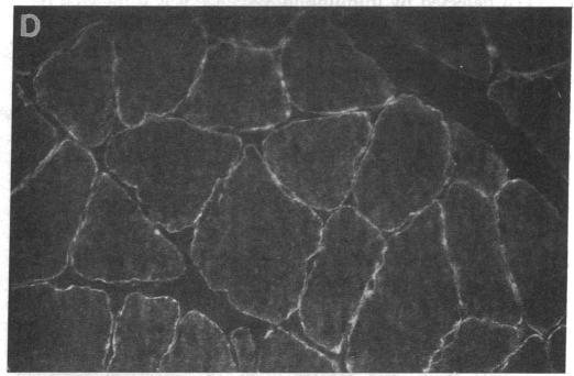
This study was designed to determine whether altered glucose transporter expression is essential for the in vivo insulin-resistant glucose uptake characteristic of streptozocin-induced diabetes. Immunofluorescence in rat skeletal muscle colocalizes GLUT4 with dystrophin, both intrinsic to muscle fibers. In contrast, GLUT1 is extrinsic to muscle fibers, probably in perineurial sheath. Immunoblotting shows that levels of GLUT1 and GLUT4 protein per DNA in hindlimb muscle are unaltered from control levels at 7 d of diabetes but decrease to approximately 20% of control at 14 d of diabetes. This decrease is prevented by insulin treatment. In adipose cells of 7 d diabetic rats, GLUT4 levels are depressed. Thus, GLUT4 undergoes tissue-specific regulation in response to diabetes. GLUT4 and GLUT1 mRNA levels in muscle are decreased 62-70% at both 7 and 14 d of diabetes and are restored by insulin treatment. At 7 d of diabetes, when GLUT4 protein levels in muscle are unaltered, in vivo insulin-stimulated glucose uptake measured by euglycemic clamp is 54% of control. This reflects impairment in both glycogen synthesis and glycolysis and the substrate common to these two pathways, glucose-6-phosphate, is decreased approximately 30% in muscle of diabetic rats. These findings suggest a defect early in the pathway of glucose utilization, probably at the step of glucose transport. Because GLUT1 and GLUT4 levels are unaltered at 7 d of diabetes, reduced glucose uptake in muscle probably reflects impaired glucose transporter translocation or intrinsic activity. Later, at 14 d of diabetes, GLUT1 and GLUT4 protein levels are reduced, suggesting that sequential defects may contribute to the insulin-resistant glucose transport characteristic of diabetes.
本研究旨在确定葡萄糖转运蛋白表达的改变对于链脲佐菌素诱导的糖尿病体内胰岛素抵抗性葡萄糖摄取特征是否至关重要。大鼠骨骼肌中的免疫荧光显示葡萄糖转运蛋白4(GLUT4)与肌营养不良蛋白共定位,二者均为肌纤维所固有。相比之下,葡萄糖转运蛋白1(GLUT1)位于肌纤维外部,可能存在于神经束膜中。免疫印迹显示,糖尿病7天时后肢肌肉中每单位DNA的GLUT1和GLUT4蛋白水平与对照水平无差异,但在糖尿病14天时降至对照水平的约20%。胰岛素治疗可预防这种下降。在糖尿病7天的大鼠脂肪细胞中,GLUT4水平降低。因此,GLUT4会因糖尿病而发生组织特异性调节。糖尿病7天和14天时,肌肉中GLUT4和GLUT1的mRNA水平均下降62 - 70%,胰岛素治疗可使其恢复。糖尿病7天时,肌肉中GLUT4蛋白水平未改变,但通过正常血糖钳夹法测得的体内胰岛素刺激的葡萄糖摄取量为对照的54%。这反映了糖原合成和糖酵解均受损,且这两条途径的共同底物葡萄糖-6-磷酸在糖尿病大鼠肌肉中减少了约30%。这些发现提示在葡萄糖利用途径的早期存在缺陷,可能在葡萄糖转运步骤。由于糖尿病7天时GLUT1和GLUT4水平未改变,肌肉中葡萄糖摄取减少可能反映了葡萄糖转运蛋白易位或内在活性受损。后来,在糖尿病14天时,GLUT1和GLUT4蛋白水平降低,提示相继出现的缺陷可能导致了糖尿病的胰岛素抵抗性葡萄糖转运特征。