• 文献检索
  • 文档翻译
  • 深度研究
  • 学术资讯
  • Suppr Zotero 插件Zotero 插件
  • 邀请有礼
  • 套餐&价格
  • 历史记录
应用&插件
Suppr Zotero 插件Zotero 插件浏览器插件Mac 客户端Windows 客户端微信小程序
定价
高级版会员购买积分包购买API积分包
服务
文献检索文档翻译深度研究API 文档MCP 服务
关于我们
关于 Suppr公司介绍联系我们用户协议隐私条款
关注我们

Suppr 超能文献

核心技术专利:CN118964589B侵权必究
粤ICP备2023148730 号-1Suppr @ 2026

文献检索

告别复杂PubMed语法,用中文像聊天一样搜索,搜遍4000万医学文献。AI智能推荐,让科研检索更轻松。

立即免费搜索

文件翻译

保留排版,准确专业,支持PDF/Word/PPT等文件格式,支持 12+语言互译。

免费翻译文档

深度研究

AI帮你快速写综述,25分钟生成高质量综述,智能提取关键信息,辅助科研写作。

立即免费体验

超声检查:一种中心静脉置管的新方法。

Ultrasonography: a novel approach to central venous cannulation.

作者信息

Agarwal Ankit, Singh Dinesh K, Singh Anil P

机构信息

Department of Anaesthesiology and Intensive Care, BHU, Varanasi, India.

出版信息

Indian J Crit Care Med. 2009 Oct;13(4):213-6. doi: 10.4103/0972-5229.60174.

DOI:10.4103/0972-5229.60174
PMID:20436690
原文链接:https://pmc.ncbi.nlm.nih.gov/articles/PMC2856149/
Abstract

BACKGROUND

Portable ultrasound machines are highly valuable in ICUs, where a patient's condition might not permit shifting the patient to the USG department for imaging. Traditionally central lines are put blindly using anatomical landmarks, which often result in complications such as difficulty in access, misplaced lines, pneumothorax, bleeding from inadvertent arterial punctures, etc. Ultrasonography provides "real time" imaging, i.e., the needle can be visualized entering the vein.

AIMS

We performed a study to compare USG guided central venous cannulation (CVC) and conventional anatomical landmark approach to CVC, in terms of ease of cannulation, time consumed, and associated complications.

SETTINGS AND DESIGN

The study was performed in a 16-bed open ICU. Eighty patients were randomly divided in two groups.

MATERIALS AND METHODS

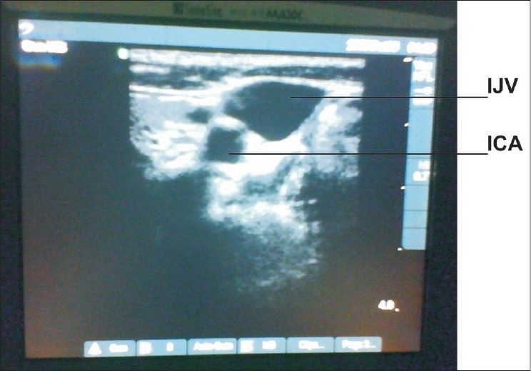
The right internal jugular vein (IJV) was cannulated in all. In Group I, a portable ultrasound machine was used during cannulation. The vessels were visualized in the transverse section with the internal carotid artery (ICA) identified as a circular pulsatile structure, while the IJV as a lateral, oval nonpulsatile structure). The needle was inserted perpendicular to the skin under visualization on the US screen. Central venous line was then inserted by the Seldinger technique. In Group II, CVC was performed by the conventional landmark approach. The parameters studied included time for insertion, attempts required, and complications encountered.

STATISTICAL ANALYSIS

The database of all parameters was analyzed using SPSS software version 10.5.

RESULTS

The mean time to successful insertion was 145 and 176.4 sec in groups I and II, respectively (p = 0.00). An average of 1.2 attempts per cannulation was required for group I, while 1.53 for group II (p = 0.03): 10% witnessed arterial puncture and 2.5% pneumothorax in group I and none in group II.

CONCLUSION

USG-guided CVC is thus easier, quicker, and safer than landmark approach.

摘要

背景

便携式超声机在重症监护病房(ICU)中具有很高的价值,因为患者的病情可能不允许将其转移到超声检查科室进行成像。传统上,中心静脉置管是盲目利用解剖标志进行的,这常常导致诸如穿刺困难、置管位置不当、气胸、意外动脉穿刺出血等并发症。超声检查可提供“实时”成像,即可以看到针头进入静脉。

目的

我们进行了一项研究,比较超声引导下中心静脉置管(CVC)和传统解剖标志法进行CVC在置管的难易程度、耗时及相关并发症方面的差异。

设置与设计

该研究在一个拥有16张床位的开放式ICU中进行。80名患者被随机分为两组。

材料与方法

所有患者均进行右颈内静脉置管。在第一组中,置管过程中使用便携式超声机。在横断面上观察血管,将颈内动脉(ICA)识别为圆形搏动结构,而颈内静脉为外侧椭圆形无搏动结构。在超声屏幕可视下将针头垂直于皮肤插入。然后采用Seldinger技术插入中心静脉导管。在第二组中,采用传统的解剖标志法进行CVC。研究的参数包括插入时间、所需尝试次数和遇到的并发症。

统计分析

使用SPSS 10.5软件对所有参数的数据库进行分析。

结果

第一组和第二组成功插入的平均时间分别为145秒和176.4秒(p = 0.00)。第一组每次置管平均需要1.2次尝试,而第二组为1.53次(p = 0.03):第一组有10%发生动脉穿刺,2.5%发生气胸,第二组均未发生。

结论

因此,超声引导下的CVC比解剖标志法更容易、更快且更安全。

https://cdn.ncbi.nlm.nih.gov/pmc/blobs/0059/2856149/f55629b362dd/IJCCM-13-213-g002.jpg
https://cdn.ncbi.nlm.nih.gov/pmc/blobs/0059/2856149/dd39f2064ec3/IJCCM-13-213-g001.jpg
https://cdn.ncbi.nlm.nih.gov/pmc/blobs/0059/2856149/f55629b362dd/IJCCM-13-213-g002.jpg
https://cdn.ncbi.nlm.nih.gov/pmc/blobs/0059/2856149/dd39f2064ec3/IJCCM-13-213-g001.jpg
https://cdn.ncbi.nlm.nih.gov/pmc/blobs/0059/2856149/f55629b362dd/IJCCM-13-213-g002.jpg

相似文献

1
Ultrasonography: a novel approach to central venous cannulation.超声检查:一种中心静脉置管的新方法。
Indian J Crit Care Med. 2009 Oct;13(4):213-6. doi: 10.4103/0972-5229.60174.
2
Incidence of posterior wall penetration during internal jugular vein cannulation: A comparison of two techniques using real-time ultrasound.颈内静脉置管时后壁穿透的发生率:两种实时超声技术的比较
Indian J Anaesth. 2017 Mar;61(3):240-244. doi: 10.4103/ija.IJA_632_16.
3
Ultrasound evaluation of the anatomical characteristics of the internal jugular vein and carotid artery--facilitation of internal jugular vein cannulation.颈内静脉和颈动脉解剖特征的超声评估——有助于颈内静脉插管
Middle East J Anaesthesiol. 2008 Oct;19(6):1305-20.
4
A Comparison of Internal Jugular Vein Cannulation by Ultrasound-Guided and Anatomical Landmark Technique in Resource-Limited Emergency Department Setting.资源有限的急诊科环境中超声引导与解剖标志技术进行颈内静脉置管的比较
J Med Ultrasound. 2019 May 13;27(4):187-191. doi: 10.4103/JMU.JMU_2_19. eCollection 2019 Oct-Dec.
5
Comparison of ultrasound guided brachiocephalic and internal jugular vein cannulation in critically ill children.危重症儿童超声引导下经头臂静脉与颈内静脉置管的比较
J Crit Care. 2016 Oct;35:133-7. doi: 10.1016/j.jcrc.2016.05.010. Epub 2016 May 25.
6
[Ultrasonic guided cannulation of the axillary vein in intensive care patients].[重症监护患者腋静脉的超声引导下置管术]
Anaesthesist. 1994 Oct;43(10):674-9. doi: 10.1007/s001010050109.
7
Comparative evaluation of success of ultrasound-guided internal jugular vein cannulation using needle with guard: A randomized, controlled study.使用带护套针头进行超声引导下颈内静脉置管成功率的比较评估:一项随机对照研究。
J Anaesthesiol Clin Pharmacol. 2020 Jan-Mar;36(1):57-61. doi: 10.4103/joacp.JOACP_305_18. Epub 2020 Feb 18.
8
Central Venous Cannulation of the Internal Jugular Vein Using Ultrasound-Guided and Anatomical Landmark Techniques.使用超声引导和解剖标志技术进行颈内静脉中心静脉置管
Anesth Pain Med. 2016 May 9;6(3):e35803. doi: 10.5812/aapm.35803. eCollection 2016 Jun.
9
Impact of ultrasonography on central venous catheter insertion in intensive care.超声检查对重症监护中中心静脉导管置入的影响。
Indian J Radiol Imaging. 2009 Jul-Sep;19(3):191-8. doi: 10.4103/0971-3026.54877.
10
Ultrasound-guided internal jugular vein access: comparison between short axis and long axis techniques.超声引导下颈内静脉穿刺:短轴与长轴技术的比较
Saudi J Kidney Dis Transpl. 2013 Jul;24(4):707-13. doi: 10.4103/1319-2442.113861.

引用本文的文献

1
Homemade phantoms improve ultrasound-guided vein cannulation confidence and procedural performance on patients.自制模型可提高超声引导下静脉置管的信心和操作技能。
Ulus Travma Acil Cerrahi Derg. 2022 Sep;28(9):1312-1316. doi: 10.14744/tjtes.2022.74712.
2
Central Venous Catheter Insertion: A Scoring System for Evaluation of Both the Procedure and the Operator (CVCI Score/Gaber Score).中心静脉导管置入:一种用于评估操作过程和操作者的评分系统(CVCI评分/加伯评分)。
Crit Care Res Pract. 2020 Nov 3;2020:8156801. doi: 10.1155/2020/8156801. eCollection 2020.
3
Recommendations on the Use of Ultrasound Guidance for Central and Peripheral Vascular Access in Adults: A Position Statement of the Society of Hospital Medicine.

本文引用的文献

1
Effectiveness of a novel training program for emergency medicine residents in ultrasound-guided insertion of central venous catheters.一种针对急诊医学住院医师的新型超声引导下中心静脉导管插入培训方案的效果
CJEM. 2009 Jul;11(4):343-8. doi: 10.1017/s1481803500011398.
2
A survey of the use of ultrasound during central venous catheterization.中心静脉置管期间超声使用情况的调查。
Anesth Analg. 2007 Mar;104(3):491-7. doi: 10.1213/01.ane.0000255289.78333.c2.
3
Real-time ultrasonographically-guided internal jugular vein catheterization in the emergency department increases success rates and reduces complications: a randomized, prospective study.
《成人中心和外周血管通路超声引导使用建议:医院医学协会立场声明》
J Hosp Med. 2019 Sep;14(9):E1-E22. doi: 10.12788/jhm.3287.
4
Cost-effectiveness-analysis of ultrasound guidance for central venous catheterization compared with landmark method: a decision-analytic model.超声引导与地标法用于中心静脉置管的成本效果分析:决策分析模型。
BMC Anesthesiol. 2019 Apr 9;19(1):51. doi: 10.1186/s12871-019-0719-5.
5
Pediatric Critical Care Medicine Training in India: Past, Present, and Future.印度的儿科重症医学培训:过去、现在与未来。
Front Pediatr. 2018 Feb 26;6:34. doi: 10.3389/fped.2018.00034. eCollection 2018.
6
Randomized controlled trials in central vascular access devices: A scoping review.中心血管通路装置的随机对照试验:一项范围综述。
PLoS One. 2017 Mar 21;12(3):e0174164. doi: 10.1371/journal.pone.0174164. eCollection 2017.
7
Evaluation of cost-effectiveness from the funding body's point of view of ultrasound-guided central venous catheter insertion compared with the conventional technique.从资助机构的角度评估超声引导下中心静脉导管插入术与传统技术相比的成本效益。
Rev Bras Ter Intensiva. 2016 Jan-Mar;28(1):62-9. doi: 10.5935/0103-507X.20160014.
8
Comparison between long- and short-axis techniques for ultrasound-guided cannulation of internal jugular vein.超声引导下颈内静脉置管的长轴与短轴技术比较
Ann Card Anaesth. 2016 Apr-Jun;19(2):288-92. doi: 10.4103/0971-9784.179629.
9
Ultrasound guidance versus anatomical landmarks for internal jugular vein catheterization.超声引导与解剖标志用于颈内静脉置管的比较
Cochrane Database Syst Rev. 2015 Jan 9;1(1):CD006962. doi: 10.1002/14651858.CD006962.pub2.
10
An entirely echo-guided technique for totally implantable access port positioning.一种用于完全植入式接入端口定位的全超声引导技术。
Indian J Surg. 2014 Jun;76(3):204-6. doi: 10.1007/s12262-012-0692-4. Epub 2012 Jul 13.
急诊科实时超声引导下颈内静脉置管可提高成功率并减少并发症:一项随机前瞻性研究。
Ann Emerg Med. 2006 Nov;48(5):540-7. doi: 10.1016/j.annemergmed.2006.01.011. Epub 2006 Feb 21.
4
Randomized, controlled clinical trial of point-of-care limited ultrasonography assistance of central venous cannulation: the Third Sonography Outcomes Assessment Program (SOAP-3) Trial.即时床旁有限超声引导下中心静脉置管的随机对照临床试验:第三次超声检查结果评估项目(SOAP-3)试验
Crit Care Med. 2005 Aug;33(8):1764-9. doi: 10.1097/01.ccm.0000171533.92856.e5.
5
A survey of the use of portable ultrasound for central vein cannulation on critical care units in the UK.
Anaesthesia. 2002 Apr;57(4):365-8. doi: 10.1046/j.1365-2044.2002.02319.x.
6
Making health care safer: a critical analysis of patient safety practices.提升医疗保健安全性:对患者安全实践的批判性分析。
Evid Rep Technol Assess (Summ). 2001(43):i-x, 1-668.
7
Facilitation of internal jugular venous cannulation using an audio-guided Doppler ultrasound vascular access device: results from a prospective, dual-center, randomized, crossover clinical study.使用音频引导多普勒超声血管通路装置促进颈内静脉置管:一项前瞻性、双中心、随机、交叉临床研究的结果
Crit Care Med. 1995 Jan;23(1):60-5. doi: 10.1097/00003246-199501000-00012.