Stanke Jennifer, Moose Holly E, El-Hodiri Heithem M, Fischer Andy J
Integrated Biomedical Science Graduate Program, College of Medicine, The Ohio State University, Columbus, Ohio 43210, USA.
J Comp Neurol. 2010 Jun 15;518(12):2316-33. doi: 10.1002/cne.22335.
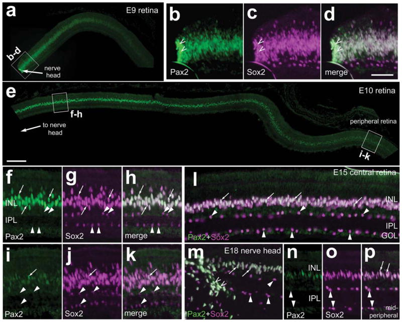
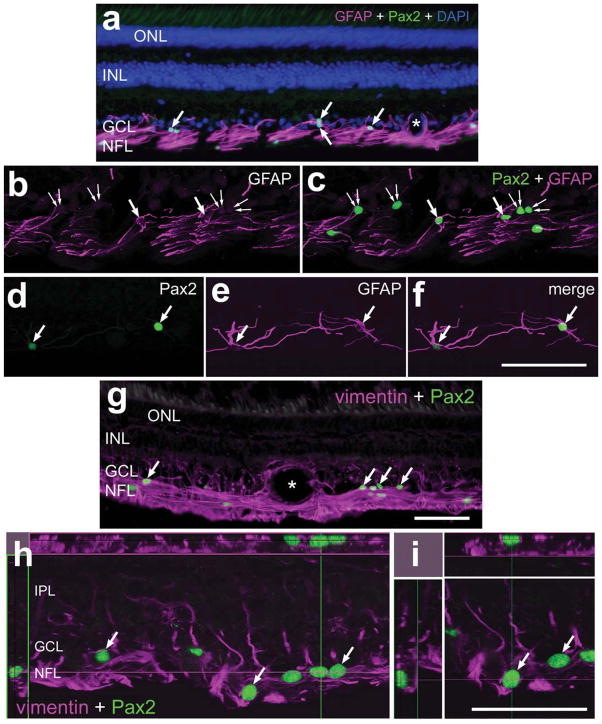
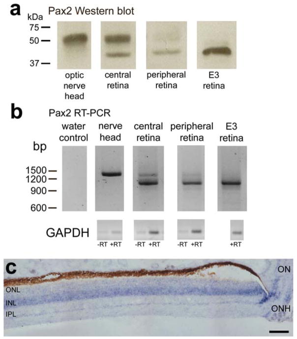
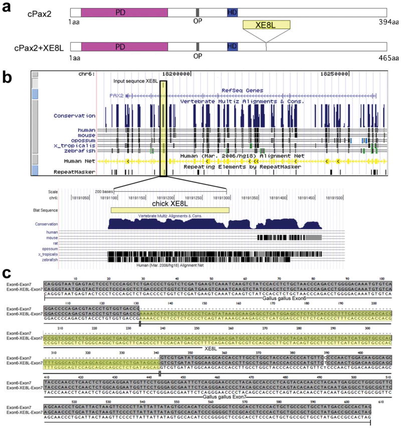
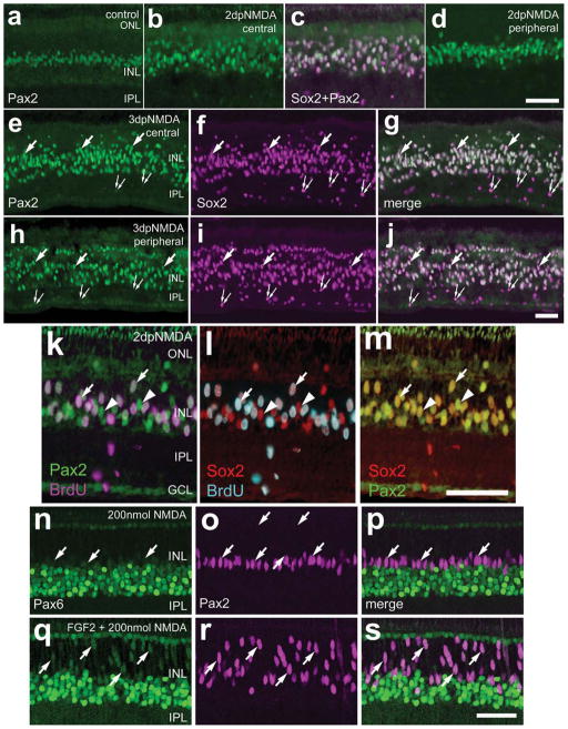
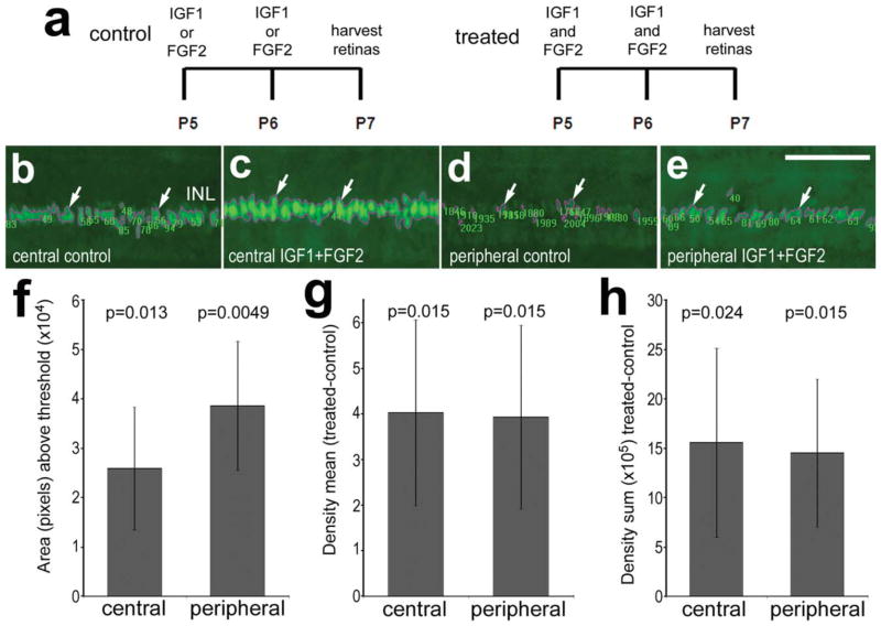
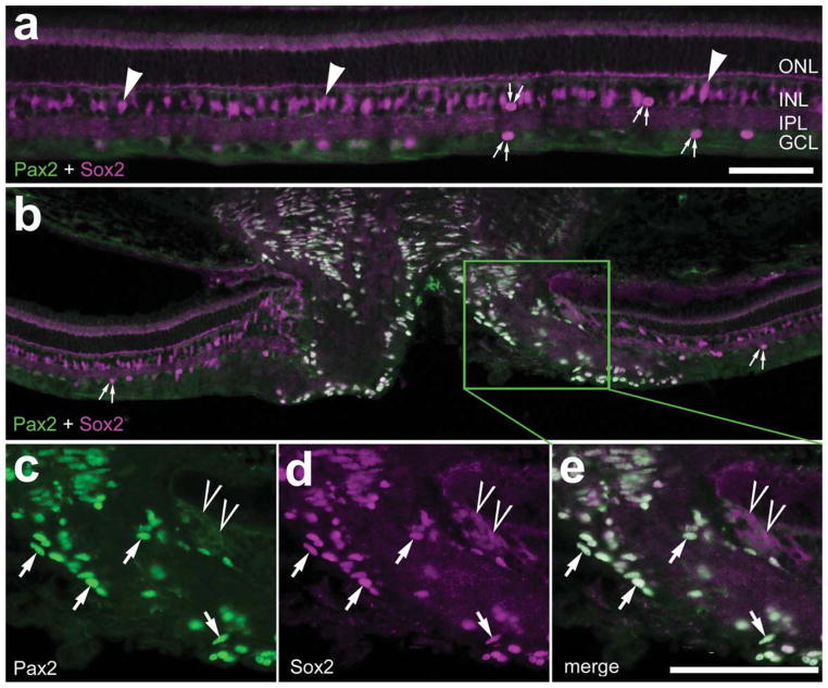
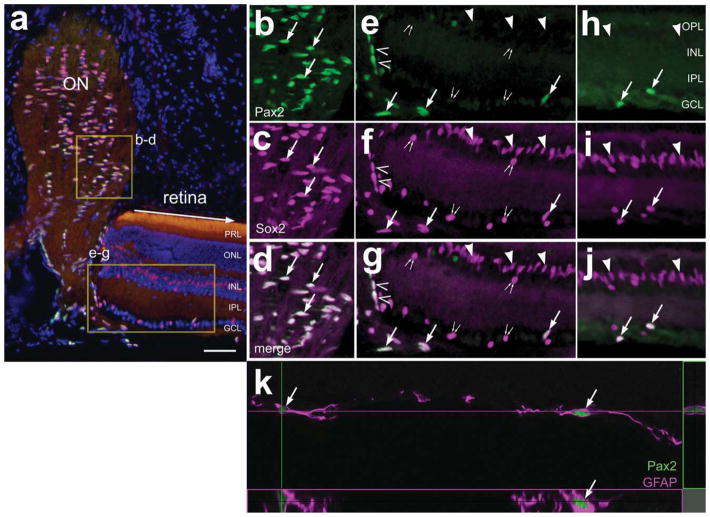
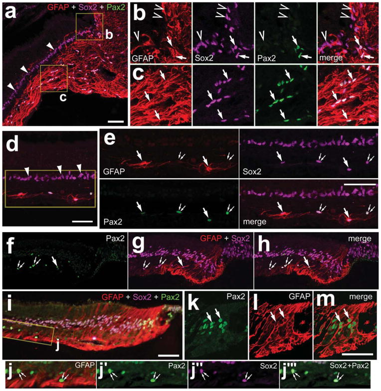
Little is known about the expression of Pax2 in mature retina or optic nerve. Here we probed for the expression of Pax2 in late stages of embryonic development and in mature chick retina. We find two distinct Pax2 isoforms expressed by cells within the retina and optic nerve. Surprisingly, Müller glia in central regions of the retina express Pax2, and levels of expression are decreased with increasing distance from the nerve head. In Müller glia, the expression levels of Pax2 are increased by acute retinal damage or treatment with growth factors. At the optic nerve, Pax2 is expressed by peripapillary glia, at the junction of the neural retina and optic nerve head and by glia within the optic nerve. In addition, we assayed for Pax2 expression in glial cells in mammalian retinas. In mammalian retinas, unlike the case in chick retina, the Müller glia do not express Pax2. Pax2-expressing cells are found in the optic nerve and astrocytes within the mouse retina. By comparison, Pax2-positive cells are not found within the guinea pig retina; Pax2-expressing glia are confined to the optic nerve. In dog and monkey (Macaca fascicularis), Pax2 is expressed by astrocytes that are scattered across inner retinal layers and by numerous glia within the optic nerve. Interestingly, Pax2-positive glial cells are found at the peripheral edge of the dog retina, but only in older animals. We conclude that the expression of Pax2 in the vertebrate eye is restricted to retinal astrocytes, peripapillary glia, and glia within the optic nerve.
关于Pax2在成熟视网膜或视神经中的表达,人们了解甚少。在此,我们探究了Pax2在胚胎发育后期和成熟鸡视网膜中的表达情况。我们发现视网膜和视神经中的细胞表达两种不同的Pax2亚型。令人惊讶的是,视网膜中央区域的穆勒胶质细胞表达Pax2,且表达水平随着与视乳头距离的增加而降低。在穆勒胶质细胞中,急性视网膜损伤或生长因子处理会使Pax2的表达水平升高。在视神经处,视乳头周围的胶质细胞、神经视网膜与视乳头头部的交界处以及视神经内的胶质细胞均表达Pax2。此外,我们检测了哺乳动物视网膜胶质细胞中Pax2的表达。在哺乳动物视网膜中,与鸡视网膜不同,穆勒胶质细胞不表达Pax2。在小鼠视网膜的视神经和星形胶质细胞中发现了表达Pax2的细胞。相比之下,在豚鼠视网膜中未发现Pax2阳性细胞;表达Pax2的胶质细胞局限于视神经。在狗和猕猴(食蟹猴)中,Pax2由散布在内层视网膜各层的星形胶质细胞以及视神经内的许多胶质细胞表达。有趣的是,在狗视网膜的周边边缘发现了Pax2阳性胶质细胞,但仅在年龄较大的动物中存在。我们得出结论,脊椎动物眼中Pax2的表达仅限于视网膜星形胶质细胞、视乳头周围的胶质细胞以及视神经内的胶质细胞。