• 文献检索
  • 文档翻译
  • 深度研究
  • 学术资讯
  • Suppr Zotero 插件Zotero 插件
  • 邀请有礼
  • 套餐&价格
  • 历史记录
应用&插件
Suppr Zotero 插件Zotero 插件浏览器插件Mac 客户端Windows 客户端微信小程序
定价
高级版会员购买积分包购买API积分包
服务
文献检索文档翻译深度研究API 文档MCP 服务
关于我们
关于 Suppr公司介绍联系我们用户协议隐私条款
关注我们

Suppr 超能文献

核心技术专利:CN118964589B侵权必究
粤ICP备2023148730 号-1Suppr @ 2026

文献检索

告别复杂PubMed语法,用中文像聊天一样搜索,搜遍4000万医学文献。AI智能推荐,让科研检索更轻松。

立即免费搜索

文件翻译

保留排版,准确专业,支持PDF/Word/PPT等文件格式,支持 12+语言互译。

免费翻译文档

深度研究

AI帮你快速写综述,25分钟生成高质量综述,智能提取关键信息,辅助科研写作。

立即免费体验

CT中的心室边界:部分容积效应与局部阈值

Ventricle Boundary in CT: Partial Volume Effect and Local Thresholds.

作者信息

Volkau Ihar, Puspitasari Fiftarina, Nowinski Wieslaw L

机构信息

Biomedical Imaging Laboratory, Agency for Science, Technology and Research (ASTAR), 30 Biopolis Street, #07-01, Matrix, Singapore 138671.

出版信息

Int J Biomed Imaging. 2010;2010:674582. doi: 10.1155/2010/674582. Epub 2010 May 17.

DOI:10.1155/2010/674582
PMID:20490355
原文链接:https://pmc.ncbi.nlm.nih.gov/articles/PMC2872763/
Abstract

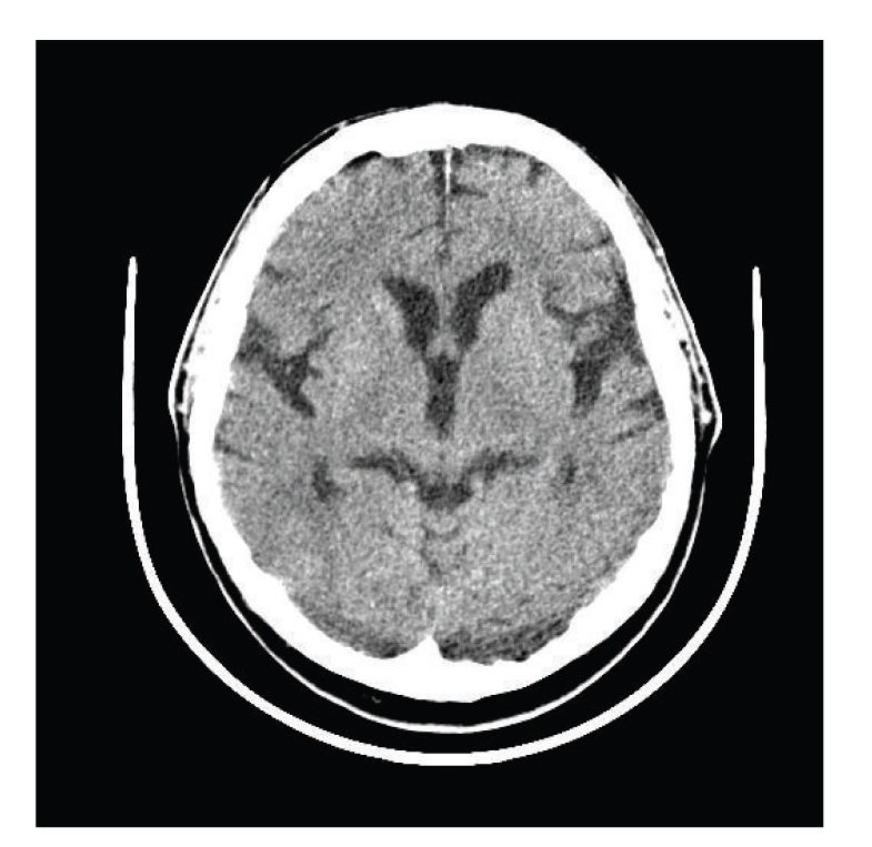
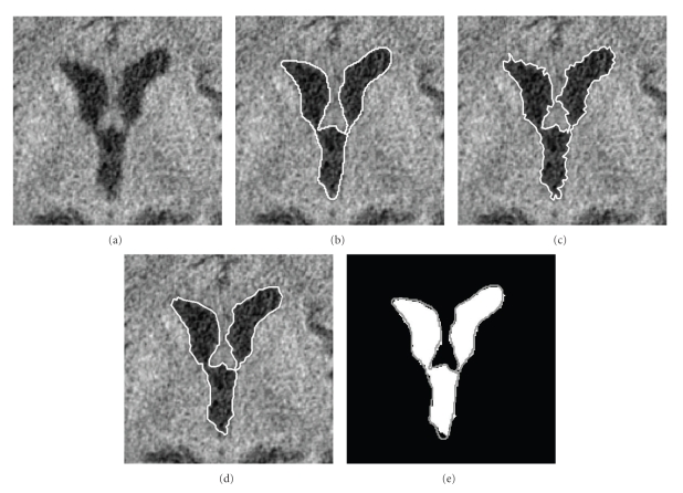
We present a mathematical frame to carry out segmentation of cerebrospinal fluid (CSF) of ventricular region in computed tomography (CT) images in the presence of partial volume effect (PVE). First, the image histogram is fitted using the Gaussian mixture model (GMM). Analyzing the GMM, we find global threshold based on parameters of distributions for CSF, and for the combined white and grey matter (WGM). The parameters of distribution of PVE pixels on the boundary of ventricles are estimated by using a convolution operator. These parameters are used to calculate local thresholds for boundary pixels by the analysis of contribution of the neighbor pixels intensities into a PVE pixel. The method works even in the case of an almost unimodal histogram; it can be useful to analyze the parameters of PVE in the ground truth provided by the expert.

摘要

我们提出了一个数学框架,用于在存在部分容积效应(PVE)的情况下,对计算机断层扫描(CT)图像中的脑室区域脑脊液(CSF)进行分割。首先,使用高斯混合模型(GMM)拟合图像直方图。通过分析GMM,我们基于CSF以及白质和灰质组合(WGM)的分布参数找到全局阈值。使用卷积算子估计脑室边界上PVE像素的分布参数。通过分析相邻像素强度对PVE像素的贡献,利用这些参数计算边界像素的局部阈值。即使在直方图几乎为单峰的情况下,该方法也能起作用;它有助于分析专家提供的真实数据中PVE的参数。

https://cdn.ncbi.nlm.nih.gov/pmc/blobs/1620/2872763/99183579a548/IJBI2010-674582.003.jpg
https://cdn.ncbi.nlm.nih.gov/pmc/blobs/1620/2872763/61974eac8dc7/IJBI2010-674582.001.jpg
https://cdn.ncbi.nlm.nih.gov/pmc/blobs/1620/2872763/298737ee67f3/IJBI2010-674582.002.jpg
https://cdn.ncbi.nlm.nih.gov/pmc/blobs/1620/2872763/99183579a548/IJBI2010-674582.003.jpg
https://cdn.ncbi.nlm.nih.gov/pmc/blobs/1620/2872763/61974eac8dc7/IJBI2010-674582.001.jpg
https://cdn.ncbi.nlm.nih.gov/pmc/blobs/1620/2872763/298737ee67f3/IJBI2010-674582.002.jpg
https://cdn.ncbi.nlm.nih.gov/pmc/blobs/1620/2872763/99183579a548/IJBI2010-674582.003.jpg

相似文献

1
Ventricle Boundary in CT: Partial Volume Effect and Local Thresholds.CT中的心室边界:部分容积效应与局部阈值
Int J Biomed Imaging. 2010;2010:674582. doi: 10.1155/2010/674582. Epub 2010 May 17.
2
Methods in quantitative image analysis.定量图像分析方法。
Histochem Cell Biol. 1996 May;105(5):333-55. doi: 10.1007/BF01463655.
3
An extension Gaussian mixture model for brain MRI segmentation.一种用于脑部磁共振成像分割的扩展高斯混合模型。
Annu Int Conf IEEE Eng Med Biol Soc. 2014;2014:4711-4. doi: 10.1109/EMBC.2014.6944676.
4
Efficient Johnson-S Mixture Model for Segmentation of CT Liver Image.高效的 Johnson-S 混合模型在 CT 肝脏图像分割中的应用。
J Healthc Eng. 2022 Apr 14;2022:5654424. doi: 10.1155/2022/5654424. eCollection 2022.
5
Positron emission tomography metabolic data corrected for cortical atrophy using magnetic resonance imaging.使用磁共振成像对皮质萎缩进行校正后的正电子发射断层扫描代谢数据。
Alzheimer Dis Assoc Disord. 1996 Fall;10(3):141-70. doi: 10.1097/00002093-199601030-00005.
6
Automatic segmentation of cerebrospinal fluid, white and gray matter in unenhanced computed tomography images.脑实质、脑白质和脑脊髓液在未增强 CT 图像中的自动分割。
Acad Radiol. 2010 Nov;17(11):1350-8. doi: 10.1016/j.acra.2010.06.005. Epub 2010 Jul 15.
7
Segmentation of SEM images of multiphase materials: When Gaussian mixture models are accurate?多相材料扫描电子显微镜图像的分割:高斯混合模型何时准确?
J Microsc. 2023 Jan;289(1):58-70. doi: 10.1111/jmi.13150. Epub 2022 Oct 27.
8
Differentiation of adrenal adenomas from nonadenomas using CT histogram analysis method: a prospective study.应用 CT 直方图分析方法对肾上腺腺瘤与非腺瘤进行鉴别诊断:一项前瞻性研究。
Eur J Radiol. 2010 Mar;73(3):643-51. doi: 10.1016/j.ejrad.2008.12.010. Epub 2009 Jan 22.
9
The calculation of CSF spaces in CT.
Neuroradiology. 1978;16:190-2. doi: 10.1007/BF00395247.
10
Partial volume correction and image segmentation for accurate measurement of standardized uptake value of grey matter in the brain.用于准确测量大脑灰质标准化摄取值的部分容积校正和图像分割
Nucl Med Commun. 2015 Dec;36(12):1249-52. doi: 10.1097/MNM.0000000000000394.

引用本文的文献

1
Quantitative estimation of a ratio of intracranial cerebrospinal fluid volume to brain volume based on segmentation of CT images in patients with extra-axial hematoma.基于轴外血肿患者CT图像分割的颅内脑脊液体积与脑体积比值的定量估计。
Neuroradiol J. 2017 Feb;30(1):10-14. doi: 10.1177/1971400916678227. Epub 2016 Nov 11.
2
Validated automatic brain extraction of head CT images.头部CT图像的经验证的自动脑提取
Neuroimage. 2015 Jul 1;114:379-85. doi: 10.1016/j.neuroimage.2015.03.074. Epub 2015 Apr 7.
3
Automated delineation of stroke lesions using brain CT images.

本文引用的文献

1
Automatic segmentation of the human brain ventricles from MR images by knowledge-based region growing and trimming.基于知识的区域生长与修剪法对磁共振图像中的人脑脑室进行自动分割
Neuroinformatics. 2009 Jun;7(2):131-46. doi: 10.1007/s12021-009-9046-1. Epub 2009 May 16.
2
Segmentation of brain from computed tomography head images.从计算机断层扫描头部图像中分割出大脑。
Conf Proc IEEE Eng Med Biol Soc. 2005;2005:3375-8. doi: 10.1109/IEMBS.2005.1617201.
3
A knowledge-driven algorithm for a rapid and automatic extraction of the human cerebral ventricular system from MR neuroimages.
利用脑部CT图像自动勾勒中风病灶
Neuroimage Clin. 2014 Mar 21;4:540-8. doi: 10.1016/j.nicl.2014.03.009. eCollection 2014.
一种基于知识的算法,用于从磁共振神经影像中快速自动提取人类脑室系统。
Neuroimage. 2004 Jan;21(1):269-82. doi: 10.1016/j.neuroimage.2003.09.029.
4
Automatic segmentation of the ventricular system from MR images of the human brain.从人脑的磁共振图像中自动分割脑室系统。
Neuroimage. 2001 Jul;14(1 Pt 1):95-104. doi: 10.1006/nimg.2001.0800.
5
An adaptive-focus statistical shape model for segmentation and shape modeling of 3-D brain structures.一种用于三维脑结构分割和形状建模的自适应聚焦统计形状模型。
IEEE Trans Med Imaging. 2001 Apr;20(4):257-70. doi: 10.1109/42.921475.
6
Precise segmentation of the lateral ventricles and caudate nucleus in MR brain images using anatomically driven histograms.利用解剖学驱动直方图对脑部磁共振图像中的侧脑室和尾状核进行精确分割。
IEEE Trans Med Imaging. 1998 Apr;17(2):303-10. doi: 10.1109/42.700743.
7
MR-based brain and cerebrospinal fluid measurement after traumatic brain injury: correlation with neuropsychological outcome.创伤性脑损伤后基于磁共振成像的脑和脑脊液测量:与神经心理学结果的相关性
AJNR Am J Neuroradiol. 1997 Jan;18(1):1-10.
8
Automated grading of venous beading.静脉串珠样改变的自动分级
Comput Biomed Res. 1995 Aug;28(4):291-304. doi: 10.1006/cbmr.1995.1020.
9
The analysis of cell images.细胞图像分析。
Ann N Y Acad Sci. 1966 Jan 31;128(3):1035-53. doi: 10.1111/j.1749-6632.1965.tb11715.x.