INSERM UMR915, l'institut du thorax, IRT-Université de Nantes, 8 quai Moncousu, BP 70721, Nantes, F-44007 Cedex 1, France.
Stem Cell Res Ther. 2010 Mar 15;1(1):4. doi: 10.1186/scrt4.
Early randomized clinical trials of autologous bone marrow cardiac stem cell therapy have reported contradictory results highlighting the need for a better evaluation of protocol designs. This study was designed to quantify and compare whole body and heart cell distribution after intracoronary or peripheral intravenous injection of autologous bone marrow mononuclear cells in a porcine acute myocardial infarction model with late reperfusion.
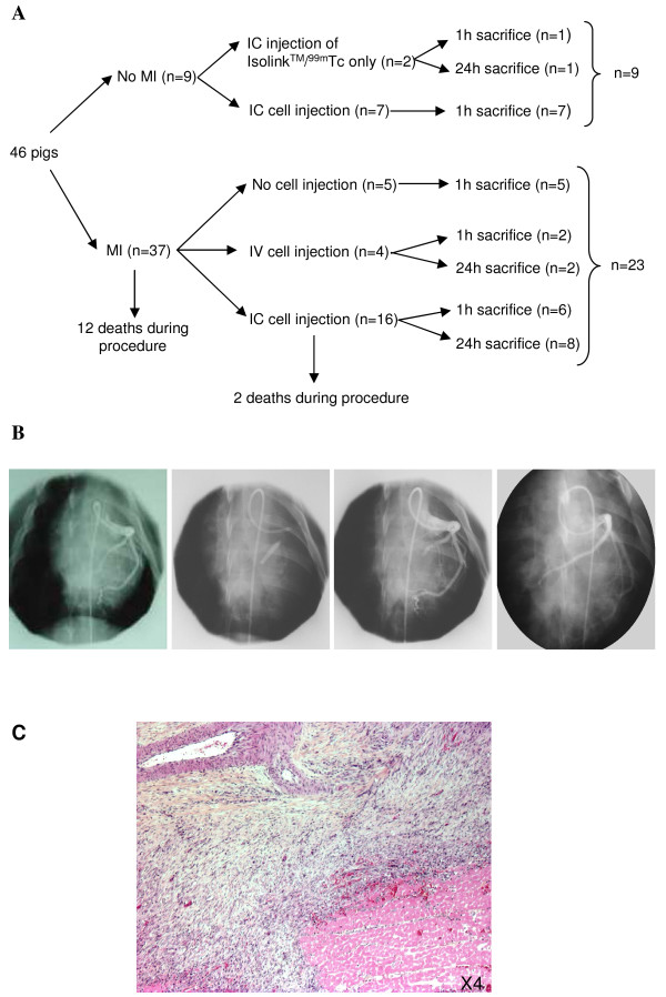
Myocardial infarction was induced using balloon inflation in the left coronary artery in domestic pigs. At seven days post-myocardial infarction, 1 x 10(8) autologous bone marrow mononuclear cells were labeled with fluorescent marker and/or 99mTc radiotracer, and delivered using intracoronary or peripheral intravenous injection (leg vein).
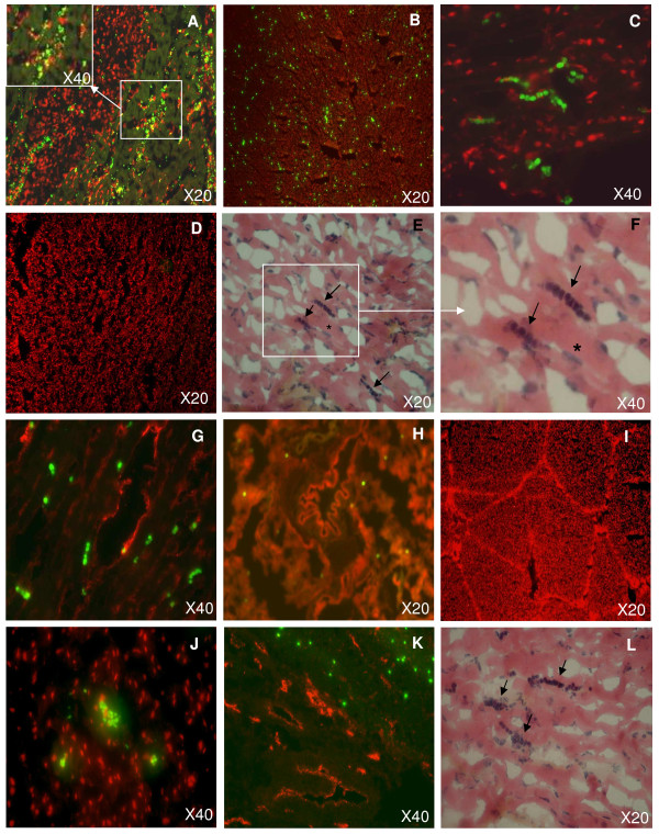
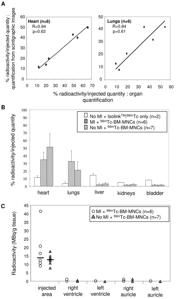
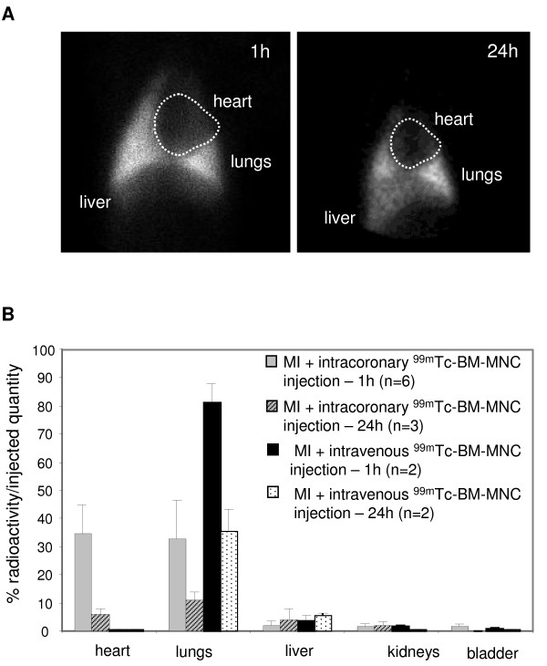
Scintigraphic analyses and Upsilon-emission radioactivity counting of harvested organs showed a significant cell fraction retained within the heart after intracoronary injection (6 +/- 1.7% of injected radioactivity at 24 hours), whereas following peripheral intravenous cell injection, no cardiac homing was observed at 24 hours and cells were mainly detected within the lungs. Importantly, no difference was observed in the percentage of retained cells within the myocardium in the presence or absence of myocardial infarction. Histological evaluation did not show arterial occlusion in both animal groups and confirmed the presence of bone marrow mononuclear cells within the injected myocardium area.
Intravenous bone marrow mononuclear cell injection was ineffective to target myocardium. Myocardial cell distribution following intracoronary injection did not depend on myocardial infarction presence, a factor that could be useful for cardiac cell therapy in patients with chronic heart failure of non-ischemic origin or with ischemic myocardium without myocardial infarction.
自体骨髓心脏干细胞治疗的早期随机临床试验结果相互矛盾,这突出表明需要更好地评估方案设计。本研究旨在通过猪急性心肌梗死模型和晚期再灌注,定量和比较经冠状动脉内或外周静脉注射自体骨髓单个核细胞后全身和心脏细胞的分布。
采用球囊在左冠状动脉内膨胀诱导心肌梗死。在心肌梗死后 7 天,将 1x10(8)个自体骨髓单个核细胞用荧光标记物和/或 99mTc 放射性示踪剂标记,并通过冠状动脉内或外周静脉注射(腿静脉)给药。
放射性核素扫描分析和采集器官的Upsilon 放射性计数显示,经冠状动脉内注射后心脏内保留了显著的细胞分数(24 小时时为注射放射性的 6+/-1.7%),而外周静脉内细胞注射后,24 小时时未观察到心脏归巢,细胞主要在肺部检测到。重要的是,在存在或不存在心肌梗死的情况下,心肌内保留的细胞百分比没有差异。组织学评估显示两组动物的动脉均未闭塞,并证实注射心肌区域内存在骨髓单个核细胞。
静脉内骨髓单个核细胞注射不能靶向心肌。经冠状动脉内注射后心肌细胞的分布不依赖于心肌梗死的存在,这一因素对于非缺血性心力衰竭或无心肌梗死的缺血性心肌患者的心脏细胞治疗可能有用。