• 文献检索
  • 文档翻译
  • 深度研究
  • 学术资讯
  • Suppr Zotero 插件Zotero 插件
  • 邀请有礼
  • 套餐&价格
  • 历史记录
应用&插件
Suppr Zotero 插件Zotero 插件浏览器插件Mac 客户端Windows 客户端微信小程序
定价
高级版会员购买积分包购买API积分包
服务
文献检索文档翻译深度研究API 文档MCP 服务
关于我们
关于 Suppr公司介绍联系我们用户协议隐私条款
关注我们

Suppr 超能文献

核心技术专利:CN118964589B侵权必究
粤ICP备2023148730 号-1Suppr @ 2026

文献检索

告别复杂PubMed语法,用中文像聊天一样搜索,搜遍4000万医学文献。AI智能推荐,让科研检索更轻松。

立即免费搜索

文件翻译

保留排版,准确专业,支持PDF/Word/PPT等文件格式,支持 12+语言互译。

免费翻译文档

深度研究

AI帮你快速写综述,25分钟生成高质量综述,智能提取关键信息,辅助科研写作。

立即免费体验

心脏磁共振伴有水肿成像可识别有风险的心肌,并预测非 ST 段抬高型急性冠状动脉综合征患者的预后更差。

Cardiac magnetic resonance with edema imaging identifies myocardium at risk and predicts worse outcome in patients with non-ST-segment elevation acute coronary syndrome.

机构信息

Ohio State University, Columbus, Ohio 43210, USA.

出版信息

J Am Coll Cardiol. 2010 Jun 1;55(22):2480-8. doi: 10.1016/j.jacc.2010.01.047.

DOI:10.1016/j.jacc.2010.01.047
PMID:20510215
原文链接:https://pmc.ncbi.nlm.nih.gov/articles/PMC3675879/
Abstract

OBJECTIVES

The aim of this study was to define the prevalence and significance of myocardial edema in patients with non-ST-segment elevation acute coronary syndrome (NSTE-ACS).

BACKGROUND

Most patients with NSTE-ACS undergo angiography, yet not all have obstructive coronary artery disease (CAD) requiring revascularization. Identifying patients with myocardium at risk could enhance the effectiveness of an early invasive strategy. Cardiac magnetic resonance (CMR) can demonstrate edematous myocardium subjected to ischemia but has not been used to evaluate NSTE-ACS patients.

METHODS

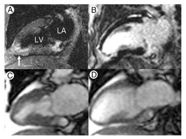
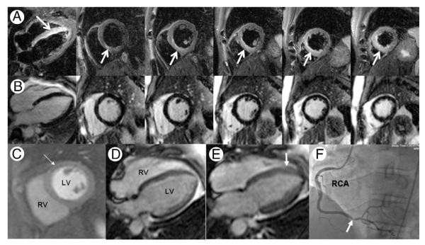
One hundred consecutive patients with NSTE-ACS were prospectively enrolled to undergo 30-min CMR, including T2-weighted edema imaging and late gadolinium enhancement before coronary angiography. Clinical management including revascularization decision-making was performed without CMR results.

RESULTS

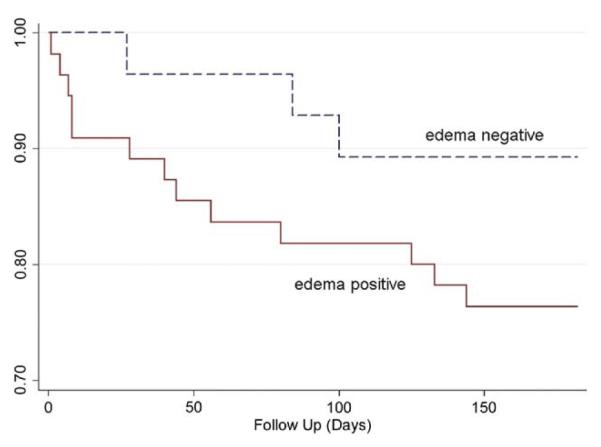
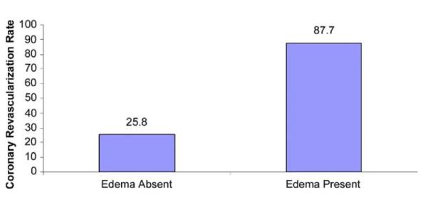
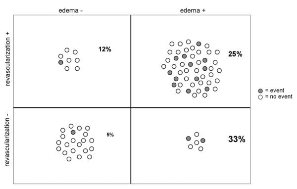
Of 88 adequate CMR studies, 57 (64.8%) showed myocardial edema. Obstructive CAD requiring revascularization was present in 87.7% of edema-positive patients versus 25.8% of edema-negative patients (p < 0.001). By multiple logistic regression analysis after adjusting for late gadolinium enhancement, perfusion, and wall motion scores, TIMI risk score was not predictive of obstructive CAD. Conversely, an increase in T2 score by 1 U increased the odds of subsequent coronary revascularization by 5.70 times (95% confidence interval: 2.38 to 13.62, p < 0.001). Adjusting for peak troponin-I, patients with edema showed a higher hazard of a cardiovascular event or death within 6 months after NSTE-ACS compared with those without edema (hazard ratio: 4.47, 95% confidence interval: 1.00 to 20.03; p = 0.050).

CONCLUSIONS

In NSTE-ACS patients, rapid CMR identifies reversibly injured myocardium due to obstructive CAD and predicts worse outcomes. Identifying myocardium at risk may help direct appropriate patients toward early invasive management.

摘要

目的

本研究旨在定义非 ST 段抬高型急性冠状动脉综合征(NSTE-ACS)患者心肌水肿的患病率和意义。

背景

大多数 NSTE-ACS 患者接受血管造影,但并非所有患者都存在需要血运重建的阻塞性冠状动脉疾病(CAD)。识别有心肌风险的患者可以提高早期侵入性策略的效果。心脏磁共振(CMR)可显示缺血导致的水肿性心肌,但尚未用于评估 NSTE-ACS 患者。

方法

前瞻性纳入 100 例 NSTE-ACS 患者,行 30 分钟 CMR,包括 T2 加权水肿成像和血管造影前晚期钆增强。临床管理包括血运重建决策,而不考虑 CMR 结果。

结果

在 88 例充分 CMR 研究中,57 例(64.8%)显示心肌水肿。在水肿阳性患者中,需要血运重建的阻塞性 CAD 占 87.7%,而在水肿阴性患者中占 25.8%(p<0.001)。经调整晚期钆增强、灌注和壁运动评分后的多变量逻辑回归分析,TIMI 风险评分不能预测阻塞性 CAD。相反,T2 评分增加 1 U,随后进行冠状动脉血运重建的几率增加 5.70 倍(95%置信区间:2.38 至 13.62,p<0.001)。调整峰值肌钙蛋白 I 后,与无水肿患者相比,有水肿的患者在 NSTE-ACS 后 6 个月内发生心血管事件或死亡的风险更高(危险比:4.47,95%置信区间:1.00 至 20.03;p=0.050)。

结论

在 NSTE-ACS 患者中,快速 CMR 可识别因阻塞性 CAD 导致的可逆性损伤心肌,并预测更差的结局。识别有风险的心肌可能有助于指导适当的患者进行早期侵入性治疗。

https://cdn.ncbi.nlm.nih.gov/pmc/blobs/04f2/3675879/f131f28f7407/nihms-475615-f0005.jpg
https://cdn.ncbi.nlm.nih.gov/pmc/blobs/04f2/3675879/a2100c288a9e/nihms-475615-f0001.jpg
https://cdn.ncbi.nlm.nih.gov/pmc/blobs/04f2/3675879/666e0a760b24/nihms-475615-f0002.jpg
https://cdn.ncbi.nlm.nih.gov/pmc/blobs/04f2/3675879/6f5ebc8712bb/nihms-475615-f0003.jpg
https://cdn.ncbi.nlm.nih.gov/pmc/blobs/04f2/3675879/2189b0af8932/nihms-475615-f0004.jpg
https://cdn.ncbi.nlm.nih.gov/pmc/blobs/04f2/3675879/f131f28f7407/nihms-475615-f0005.jpg

相似文献

1
Cardiac magnetic resonance with edema imaging identifies myocardium at risk and predicts worse outcome in patients with non-ST-segment elevation acute coronary syndrome.心脏磁共振伴有水肿成像可识别有风险的心肌,并预测非 ST 段抬高型急性冠状动脉综合征患者的预后更差。
J Am Coll Cardiol. 2010 Jun 1;55(22):2480-8. doi: 10.1016/j.jacc.2010.01.047.
2
Approach to non-ST-segment elevation acute coronary syndrome in the emergency department: risk stratification and treatment strategies.急诊科非ST段抬高型急性冠状动脉综合征的处理:风险分层与治疗策略
Hosp Pract (1995). 2010 Apr;38(2):40-9.
3
Management patterns of non-ST segment elevation acute coronary syndromes in relation to prior coronary revascularization.非 ST 段抬高型急性冠状动脉综合征与既往冠状动脉血运重建相关的管理模式。
Am Heart J. 2010 Jan;159(1):40-6. doi: 10.1016/j.ahj.2009.09.019.
4
Temporal trends in the use of invasive cardiac procedures for non-ST segment elevation acute coronary syndromes according to initial risk stratification.根据初始风险分层,非ST段抬高型急性冠状动脉综合征患者侵入性心脏手术使用情况的时间趋势。
Can J Cardiol. 2009 Nov;25(11):e370-6. doi: 10.1016/s0828-282x(09)70163-1.
5
Timing of angiography with a routine invasive strategy and long-term outcomes in non-ST-segment elevation acute coronary syndrome: a collaborative analysis of individual patient data from the FRISC II (Fragmin and Fast Revascularization During Instability in Coronary Artery Disease), ICTUS (Invasive Versus Conservative Treatment in Unstable Coronary Syndromes), and RITA-3 (Intervention Versus Conservative Treatment Strategy in Patients With Unstable Angina or Non-ST Elevation Myocardial Infarction) Trials.常规有创策略下的血管造影时机与非 ST 段抬高型急性冠脉综合征的长期预后:FRISC II(不稳定型冠状动脉疾病时的磺达肝癸钠和早期血运重建)、ICTUS(不稳定型冠状动脉综合征的有创与保守治疗)和 RITA-3(不稳定型心绞痛或非 ST 段抬高型心肌梗死患者介入与保守治疗策略)这三项试验的个体化患者数据的协作分析。
JACC Cardiovasc Interv. 2012 Feb;5(2):191-9. doi: 10.1016/j.jcin.2011.10.016.
6
Prevalence, predictors, and impact of conservative medical management for patients with non-ST-segment elevation acute coronary syndromes who have angiographically documented significant coronary disease.对于血管造影显示有明显冠状动脉疾病的非ST段抬高型急性冠状动脉综合征患者,保守药物治疗的患病率、预测因素及影响
JACC Cardiovasc Interv. 2008 Aug;1(4):369-78. doi: 10.1016/j.jcin.2008.03.019.
7
A risk score to predict bleeding in patients with acute coronary syndromes.用于预测急性冠脉综合征患者出血风险的评分。
J Am Coll Cardiol. 2010 Jun 8;55(23):2556-66. doi: 10.1016/j.jacc.2009.09.076.
8
Early invasive strategy in high-risk acute coronary syndrome without ST-segment elevation. The Sisca randomized trial.非ST段抬高型高危急性冠状动脉综合征的早期侵入性策略。Sisca随机试验。
Int J Cardiol. 2015 Mar 1;182:414-8. doi: 10.1016/j.ijcard.2014.12.089. Epub 2014 Dec 27.
9
Usefulness of layer-specific strain for identifying complex CAD and predicting the severity of coronary lesions in patients with non-ST-segment elevation acute coronary syndrome: Compared with Syntax score.非ST段抬高型急性冠状动脉综合征患者中,层特异性应变在识别复杂冠状动脉疾病和预测冠状动脉病变严重程度方面的效用:与Syntax评分的比较
Int J Cardiol. 2016 Nov 15;223:1045-1052. doi: 10.1016/j.ijcard.2016.08.277. Epub 2016 Aug 18.
10
Early Invasive Versus Selective Strategy for Non-ST-Segment Elevation Acute Coronary Syndrome: The ICTUS Trial.早期侵入性与选择性策略治疗非 ST 段抬高急性冠状动脉综合征:ICTUS 试验。
J Am Coll Cardiol. 2017 Apr 18;69(15):1883-1893. doi: 10.1016/j.jacc.2017.02.023.

引用本文的文献

1
Cardiovascular Magnetic Resonance from Fetal to Adult Life-Indications and Challenges: A State-of-the-Art Review.从胎儿期到成年期的心血管磁共振成像:适应证与挑战——一项前沿综述
Children (Basel). 2023 Apr 23;10(5):763. doi: 10.3390/children10050763.
2
Value of Fast MVO Identification From Contrast-Enhanced Cine (CE-SSFP) Combined With Myocardial Strain in Predicting Adverse Events in Patients After ST-Elevation Myocardial Infarction.从对比增强电影(CE-SSFP)结合心肌应变快速识别心肌微血管阻塞在预测ST段抬高型心肌梗死患者不良事件中的价值。
Front Cardiovasc Med. 2022 Feb 21;8:804020. doi: 10.3389/fcvm.2021.804020. eCollection 2021.
3

本文引用的文献

1
T2 quantification for improved detection of myocardial edema.用于提高心肌水肿检测的 T2 定量
J Cardiovasc Magn Reson. 2009 Dec 30;11(1):56. doi: 10.1186/1532-429X-11-56.
2
Early versus delayed invasive intervention in acute coronary syndromes.急性冠状动脉综合征的早期与延迟侵入性干预
N Engl J Med. 2009 May 21;360(21):2165-75. doi: 10.1056/NEJMoa0807986.
3
Edema as a very early marker for acute myocardial ischemia: a cardiovascular magnetic resonance study.水肿作为急性心肌缺血的极早期标志物:一项心血管磁共振研究
Myocardial oedema: pathophysiological basis and implications for the failing heart.
心肌水肿:病理生理基础及其对衰竭心脏的影响。
ESC Heart Fail. 2022 Apr;9(2):958-976. doi: 10.1002/ehf2.13775. Epub 2022 Feb 11.
4
SCMR Position Paper (2020) on clinical indications for cardiovascular magnetic resonance.SCMR 立场文件(2020 年):心血管磁共振的临床适应证。
J Cardiovasc Magn Reson. 2020 Nov 9;22(1):76. doi: 10.1186/s12968-020-00682-4.
5
Clinical Importance of Myocardial T Mapping and Texture Analysis.心肌 T 映射和纹理分析的临床重要性。
Magn Reson Med Sci. 2021 Jun 1;20(2):139-151. doi: 10.2463/mrms.rev.2020-0007. Epub 2020 May 11.
6
Depression and myocardial injury in ST-segment elevation myocardial infarction: A cardiac magnetic resonance imaging study.ST段抬高型心肌梗死中的抑郁与心肌损伤:一项心脏磁共振成像研究。
World J Clin Cases. 2020 Apr 6;8(7):1232-1240. doi: 10.12998/wjcc.v8.i7.1232.
7
Cardiac MRI Endpoints in Myocardial Infarction Experimental and Clinical Trials: JACC Scientific Expert Panel.心肌梗死实验和临床研究中的心脏 MRI 终点:美国心脏病学会科学专家小组。
J Am Coll Cardiol. 2019 Jul 16;74(2):238-256. doi: 10.1016/j.jacc.2019.05.024.
8
T1 and T2 mapping in the identification of acute myocardial injury in patients with NSTEMI.T1 和 T2 映射在 NSTEMI 患者急性心肌损伤中的识别。
Radiol Med. 2018 Dec;123(12):926-934. doi: 10.1007/s11547-018-0931-2. Epub 2018 Aug 21.
9
T-Wave Abnormality as Electrocardiographic Signature of Myocardial Edema in Non-ST-Elevation Acute Coronary Syndromes.T 波异常作为非 ST 段抬高型急性冠状动脉综合征中心肌水肿的心电图特征。
J Am Heart Assoc. 2018 Jan 26;7(3):e007118. doi: 10.1161/JAHA.117.007118.
10
Correlation of Reciprocal Changes and QRS Amplitude in ECG to Left Ventricular Dysfunction, Wall Motion Score and Clinical Outcome in First Time ST Elevation Myocardial Infarction.心电图中 reciprocal 变化及 QRS 波振幅与首次 ST 段抬高型心肌梗死患者左心室功能障碍、室壁运动评分及临床结局的相关性
J Clin Diagn Res. 2017 Jul;11(7):OC04-OC08. doi: 10.7860/JCDR/2017/26021.10155. Epub 2017 Jul 1.
J Am Coll Cardiol. 2009 Apr 7;53(14):1194-201. doi: 10.1016/j.jacc.2008.10.065.
4
Early versus delayed, provisional eptifibatide in acute coronary syndromes.急性冠状动脉综合征中早期与延迟应用临时替罗非班的比较
N Engl J Med. 2009 May 21;360(21):2176-90. doi: 10.1056/NEJMoa0901316. Epub 2009 Mar 30.
5
Patterns of use and effectiveness of early invasive strategy in non-ST-segment elevation acute coronary syndromes: an assessment by propensity score.非ST段抬高型急性冠状动脉综合征早期侵入性策略的使用模式及有效性:一项倾向评分评估
Am Heart J. 2008 Nov;156(5):946-53, 953.e2. doi: 10.1016/j.ahj.2008.06.032. Epub 2008 Sep 11.
6
Diverging associations of an intended early invasive strategy compared with actual revascularization, and outcome in patients with non-ST-segment elevation acute coronary syndrome: the problem of treatment selection bias.与实际血运重建相比,非ST段抬高型急性冠脉综合征患者采用预期早期侵入性策略的不同关联及结局:治疗选择偏倚问题
Eur Heart J. 2009 Mar;30(6):645-54. doi: 10.1093/eurheartj/ehn438. Epub 2008 Sep 29.
7
Cardiac magnetic resonance with T2-weighted imaging improves detection of patients with acute coronary syndrome in the emergency department.采用T2加权成像的心脏磁共振成像可提高急诊科急性冠状动脉综合征患者的检出率。
Circulation. 2008 Aug 19;118(8):837-44. doi: 10.1161/CIRCULATIONAHA.107.740597. Epub 2008 Aug 4.
8
Early invasive vs conservative treatment strategies in women and men with unstable angina and non-ST-segment elevation myocardial infarction: a meta-analysis.不稳定型心绞痛和非ST段抬高型心肌梗死患者中早期侵入性与保守治疗策略的比较:一项荟萃分析
JAMA. 2008 Jul 2;300(1):71-80. doi: 10.1001/jama.300.1.71.
9
Mechanics of the left ventricular myocardial interstitium: effects of acute and chronic myocardial edema.左心室心肌间质力学:急性和慢性心肌水肿的影响
Am J Physiol Heart Circ Physiol. 2008 Jun;294(6):H2428-34. doi: 10.1152/ajpheart.00860.2007. Epub 2008 Mar 28.
10
Detecting patients with acute coronary syndrome in the chest pain center of the emergency department with cardiac magnetic resonance imaging.利用心脏磁共振成像在急诊科胸痛中心检测急性冠状动脉综合征患者。
Crit Pathw Cardiol. 2004 Mar;3(1):25-31. doi: 10.1097/01.hpc.0000116584.57152.06.