• 文献检索
  • 文档翻译
  • 深度研究
  • 学术资讯
  • Suppr Zotero 插件Zotero 插件
  • 邀请有礼
  • 套餐&价格
  • 历史记录
应用&插件
Suppr Zotero 插件Zotero 插件浏览器插件Mac 客户端Windows 客户端微信小程序
定价
高级版会员购买积分包购买API积分包
服务
文献检索文档翻译深度研究API 文档MCP 服务
关于我们
关于 Suppr公司介绍联系我们用户协议隐私条款
关注我们

Suppr 超能文献

核心技术专利:CN118964589B侵权必究
粤ICP备2023148730 号-1Suppr @ 2026

文献检索

告别复杂PubMed语法,用中文像聊天一样搜索,搜遍4000万医学文献。AI智能推荐,让科研检索更轻松。

立即免费搜索

文件翻译

保留排版,准确专业,支持PDF/Word/PPT等文件格式,支持 12+语言互译。

免费翻译文档

深度研究

AI帮你快速写综述,25分钟生成高质量综述,智能提取关键信息,辅助科研写作。

立即免费体验

新型金属植入技术治疗内侧距骨穹窿骨软骨缺损。尸体研究。

Novel metallic implantation technique for osteochondral defects of the medial talar dome. A cadaver study.

机构信息

Orthopedic Research Center Amsterdam, Department of Orthopedic Surgery, Academic Medical Center, University of Amsterdam, The Netherlands.

出版信息

Acta Orthop. 2010 Aug;81(4):495-502. doi: 10.3109/17453674.2010.492764.

DOI:10.3109/17453674.2010.492764
PMID:20515434
原文链接:https://pmc.ncbi.nlm.nih.gov/articles/PMC2917574/
Abstract

BACKGROUND AND PURPOSE

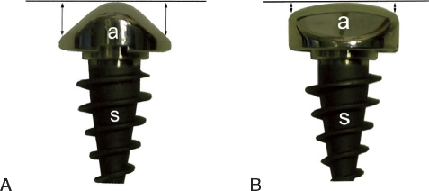
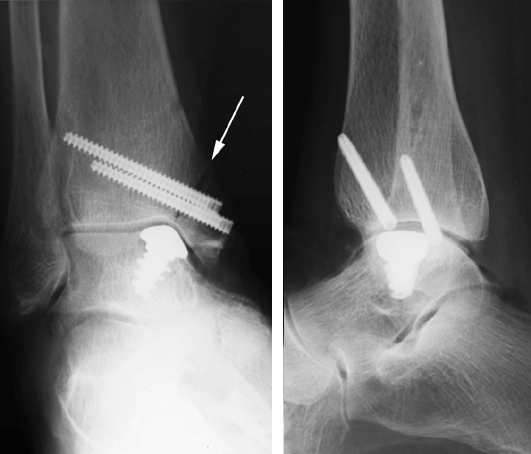
A metallic inlay implant (HemiCAP) with 15 offset sizes has been developed for the treatment of localized osteochondral defects of the medial talar dome. The aim of this study was to test the following hypotheses: (1) a matching offset size is available for each talus, (2) the prosthetic device can be reproducibly implanted slightly recessed in relation to the talar cartilage level, and (3) with this implantation level, excessive contact pressures on the opposite tibial cartilage are avoided.

METHODS

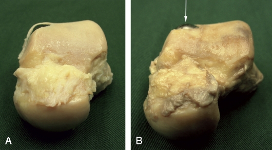
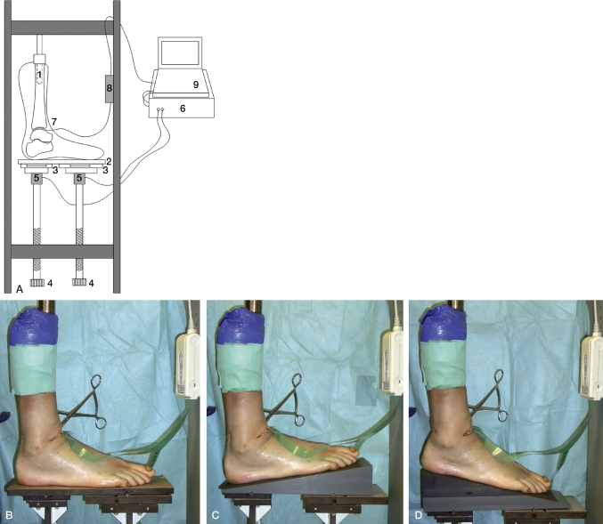
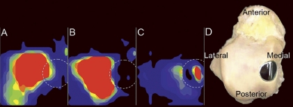
The prosthetic device was implanted in 11 intact fresh-frozen human cadaver ankles, aiming its surface 0.5 mm below cartilage level. The implantation level was measured at 4 margins of each implant. Intraarticular contact pressures were measured before and after implantation, with compressive forces of 1,000-2,000 N and the ankle joint in plantigrade position, 10 dorsiflexion, and 14 plantar flexion.

RESULTS

There was a matching offset size available for each specimen. The mean implantation level was 0.45 (SD 0.18) mm below the cartilage surface. The defect area accounted for a median of 3% (0.02-18) of the total ankle contact pressure before implantation. This was reduced to 0.1% (0.02-13) after prosthetic implantation.

INTERPRETATION

These results suggest that the implant can be applied clinically in a safe way, with appropriate offset sizes for various talar domes and without excessive pressure on the opposite cartilage.

摘要

背景与目的

为了治疗内侧距骨穹隆局灶性骨软骨缺损,开发了一种带有 15 个偏移量的金属镶嵌植入物(HemiCAP)。本研究旨在验证以下假设:(1)每个距骨都有匹配的偏移量;(2)可以将假体设备可重复地稍微凹陷植入距骨软骨平面以下;(3)在这种植入水平下,避免对相反的胫骨软骨产生过大的接触压力。

方法

将假体设备植入 11 个完整的新鲜冷冻人尸体踝关节中,目标是使其表面低于软骨平面 0.5 毫米。在每个植入物的 4 个边缘测量植入水平。在植入前和植入后,在 1,000-2,000 N 的压缩力下,踝关节处于跖屈位、10 度背屈和 14 度跖屈,测量关节内接触压力。

结果

每个标本都有匹配的偏移量。平均植入水平比软骨表面低 0.45(SD 0.18)毫米。在植入前,缺陷区域占总踝关节接触压力的中位数为 3%(0.02-18)。植入后,这一比例降至 0.1%(0.02-13)。

解释

这些结果表明,该植入物可以以安全的方式应用于临床,对于各种距骨穹隆有适当的偏移量,并且不会对相反的软骨产生过大的压力。

https://cdn.ncbi.nlm.nih.gov/pmc/blobs/67d9/2917574/fe198199e488/ORT-1745-3674-81-495-g005.jpg
https://cdn.ncbi.nlm.nih.gov/pmc/blobs/67d9/2917574/b3a837415725/ORT-1745-3674-81-495-g001.jpg
https://cdn.ncbi.nlm.nih.gov/pmc/blobs/67d9/2917574/19ccb3ba2f5c/ORT-1745-3674-81-495-g002.jpg
https://cdn.ncbi.nlm.nih.gov/pmc/blobs/67d9/2917574/009789ed5a6f/ORT-1745-3674-81-495-g003.jpg
https://cdn.ncbi.nlm.nih.gov/pmc/blobs/67d9/2917574/228fc2be4a2a/ORT-1745-3674-81-495-g004.jpg
https://cdn.ncbi.nlm.nih.gov/pmc/blobs/67d9/2917574/fe198199e488/ORT-1745-3674-81-495-g005.jpg

相似文献

1
Novel metallic implantation technique for osteochondral defects of the medial talar dome. A cadaver study.新型金属植入技术治疗内侧距骨穹窿骨软骨缺损。尸体研究。
Acta Orthop. 2010 Aug;81(4):495-502. doi: 10.3109/17453674.2010.492764.
2
Autologous osteochondral transplantation of the talus partially restores contact mechanics of the ankle joint.自体距骨骨软骨移植部分恢复踝关节的接触力学。
Am J Sports Med. 2011 Nov;39(11):2457-65. doi: 10.1177/0363546511419811. Epub 2011 Aug 25.
3
Mosaicplasty of the talus: a joint contact analysis in a cadaver model.距骨镶嵌植骨术:尸体模型中的关节接触分析
J Foot Ankle Surg. 2002 Mar-Apr;41(2):65-75. doi: 10.1016/s1067-2516(02)80030-8.
4
1994 William J. Stickel Gold Award. Contact characteristics of the ankle joint. Part 2. The effects of talar dome cartilage defects.1994年威廉·J·斯蒂克尔金奖。踝关节的接触特征。第2部分。距骨穹窿软骨缺损的影响。
J Am Podiatr Med Assoc. 1994 Nov;84(11):537-47. doi: 10.7547/87507315-84-11-537.
5
Accessibility and Thickness of Medial and Lateral Talar Body Cartilage for Treatment of Ankle and Foot Osteochondral Lesions.距骨体软骨内侧面和外侧面的可及性和厚度在治疗踝关节和足部骨软骨病变中的作用。
Foot Ankle Int. 2021 Oct;42(10):1330-1339. doi: 10.1177/10711007211015189. Epub 2021 May 28.
6
An anterior ankle arthroscopic technique for retrograde osteochondral autograft transplantation of posteromedial and central talar dome cartilage defects.一种用于后内侧和中央距骨穹顶软骨缺损逆行骨软骨自体移植的踝关节前侧关节镜技术。
Knee Surg Sports Traumatol Arthrosc. 2014 Jun;22(6):1298-303. doi: 10.1007/s00167-013-2502-3. Epub 2013 Apr 12.
7
Histomorphometric, CT arthrographic, and biomechanical mapping of the human ankle.人体踝关节的组织形态计量学、CT 关节造影和生物力学图谱。
Foot Ankle Int. 2013 Jul;34(7):1025-34. doi: 10.1177/1071100713477636. Epub 2013 Feb 8.
8
Effect of implantation accuracy on ankle contact mechanics with a metallic focal resurfacing implant.金属点状切除假体植入精度对踝关节接触力学的影响。
J Bone Joint Surg Am. 2010 Jun;92(6):1490-500. doi: 10.2106/JBJS.I.00431.
9
Kinematic changes after fusion and total replacement of the ankle: part 3: Talar movement.踝关节融合与全置换术后的运动学变化:第3部分:距骨运动
Foot Ankle Int. 2003 Dec;24(12):897-900. doi: 10.1177/107110070302401204.
10
Cartilage thickness of the talar dome.距骨穹窿的软骨厚度。
Arthroscopy. 2005 Apr;21(4):401-4. doi: 10.1016/j.arthro.2004.12.005.

引用本文的文献

1
The Frequency and Severity of Complications in Surgical Treatment of Osteochondral Lesions of the Talus: A Systematic Review and Meta-Analysis of 6,962 Lesions.距骨骨软骨病变手术治疗的并发症频率和严重程度:6962 个病灶的系统评价和荟萃分析。
Cartilage. 2023 Jun;14(2):180-197. doi: 10.1177/19476035231154746. Epub 2023 Mar 9.
2
Osteochondral Lesions of Ankle and Knee. Will Future Treatments Really Be Represented by Custom-Made Metal Implants?踝关节和膝关节的骨软骨损伤。未来的治疗真的会由定制金属植入物来代表吗?
J Clin Med. 2022 Jul 1;11(13):3817. doi: 10.3390/jcm11133817.
3
Osteochondral Lesions of the Talus: A Review on Talus Osteochondral Injuries, Including Osteochondritis Dissecans.

本文引用的文献

1
The natural history of osteochondral lesions in the ankle.踝关节骨软骨损伤的自然病史。
Instr Course Lect. 2010;59:375-86.
2
Treatment of osteochondral lesions of the talus: a systematic review.距骨骨软骨病变的治疗:系统评价。
Knee Surg Sports Traumatol Arthrosc. 2010 Feb;18(2):238-46. doi: 10.1007/s00167-009-0942-6. Epub 2009 Oct 27.
3
Donor-site morbidity after osteochondral autologous transplantation for lesions of the talus.距骨病变自体骨软骨移植术后供区并发症
距骨骨软骨病变:距骨骨软骨损伤综述,包括剥脱性骨软骨炎。
Cartilage. 2021 Dec;13(1_suppl):1380S-1401S. doi: 10.1177/1947603520985182. Epub 2021 Jan 9.
4
[Biomechanical study on repair and reconstruction of talar lesion by three-dimensional printed talar components].[三维打印距骨组件修复与重建距骨病变的生物力学研究]
Zhongguo Xiu Fu Chong Jian Wai Ke Za Zhi. 2018 Mar 15;32(3):306-310. doi: 10.7507/1002-1892.201705068.
5
Metal Resurfacing Inlay Implant for Osteochondral Talar Defects After Failed Previous Surgery: A Midterm Prospective Follow-up Study.金属表面植入物治疗前次手术失败后的距骨骨软骨缺损:中期前瞻性随访研究。
Am J Sports Med. 2018 Jun;46(7):1685-1692. doi: 10.1177/0363546518764916. Epub 2018 Apr 6.
6
Management of Osteochondral Lesions of the Talar Dome.距骨穹窿骨软骨损伤的管理
Open Orthop J. 2017 Jul 31;11:743-761. doi: 10.2174/1874325001711010743. eCollection 2017.
7
Tertiary osteochondral defect of the talus treated by a novel contoured metal implant.新型曲形金属植入物治疗距骨三级骨软骨缺损。
Knee Surg Sports Traumatol Arthrosc. 2011 Jun;19(6):999-1003. doi: 10.1007/s00167-011-1465-5. Epub 2011 Mar 16.
8
Direction of the oblique medial malleolar osteotomy for exposure of the talus.外踝斜向内侧切开术入路暴露距骨。
Arch Orthop Trauma Surg. 2011 Jul;131(7):893-901. doi: 10.1007/s00402-010-1227-8. Epub 2010 Dec 17.
J Bone Joint Surg Am. 2009 Jul;91(7):1683-8. doi: 10.2106/JBJS.H.00429.
4
Partial resurfacing with varus osteotomy for an osteochondral defect of the femoral head.采用内翻截骨术对股骨头骨软骨缺损进行部分表面重建。
Hip Int. 2009 Jan-Mar;19(1):67-70. doi: 10.1177/112070000901900113.
5
Partial humeral head resurfacing for osteonecrosis.用于治疗骨坏死的肱骨头部分表面置换术。
J Shoulder Elbow Surg. 2009 Sep-Oct;18(5):711-6. doi: 10.1016/j.jse.2008.10.016. Epub 2009 Jan 30.
6
High inter-specimen variability of baseline data for the tibio-talar contact area.胫距接触面积的基线数据在样本间存在高度变异性。
Clin Biomech (Bristol). 2009 Jan;24(1):117-20. doi: 10.1016/j.clinbiomech.2008.10.006. Epub 2008 Dec 3.
7
Treatment of osteochondral defects of the talus.距骨骨软骨损伤的治疗。
Rev Chir Orthop Reparatrice Appar Mot. 2008 Dec;94(8 Suppl):398-408. doi: 10.1016/j.rco.2008.09.003. Epub 2008 Nov 11.
8
Advancements in ankle arthroscopy.踝关节镜检查的进展。
J Am Acad Orthop Surg. 2008 Nov;16(11):635-46. doi: 10.5435/00124635-200811000-00004.
9
Focal anatomic patellofemoral inlay resurfacing: theoretic basis, surgical technique, and case reports.局灶性解剖型髌股关节镶嵌式表面置换:理论基础、手术技术及病例报告。
Orthop Clin North Am. 2008 Jul;39(3):337-46, vi. doi: 10.1016/j.ocl.2008.02.003.
10
In vivo cartilage contact deformation of human ankle joints under full body weight.人体踝关节在全身重量下的体内软骨接触变形。
J Orthop Res. 2008 Aug;26(8):1081-9. doi: 10.1002/jor.20593.