Department of Surgical Sciences, University of Wisconsin-Madison, Madison, WI, USA.
Cornea. 2010 Aug;29(8):910-6. doi: 10.1097/ICO.0b013e3181c846aa.
To assess integration of a biosynthetic corneal implant in dogs.
Three normal adult laboratory Beagles underwent ophthalmic examinations, including slit-lamp biomicroscopy, indirect ophthalmoscopy, applanation tonometry, and Cochet-Bonnet aesthesiometry. Biosynthetic corneas fabricated from glutaraldehyde crosslinked collagen and copolymers of collagen and poly(N-isopropylacrylamide-co-acrylic acid-co-acryloxysuccinimide, denoted as TERP) were implanted into dogs by a modified epikeratoplasty technique. Ophthalmic examinations and aesthesiometry were performed daily for 5 days and then weekly thereafter for 16 weeks. Corneal samples underwent histopathological and transmission electron microscopy examination at 16 weeks.
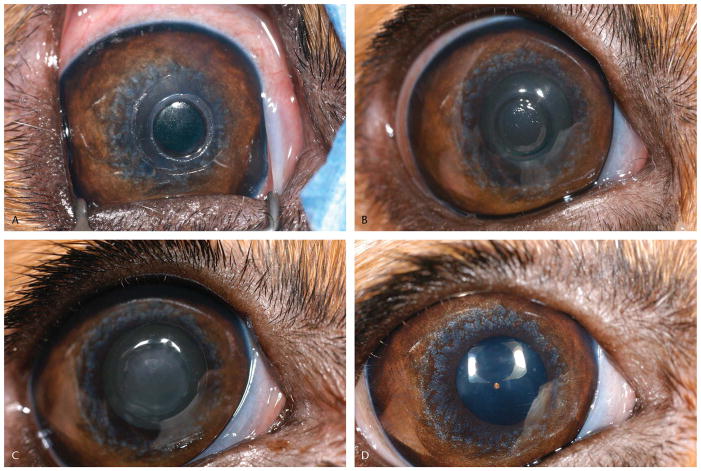
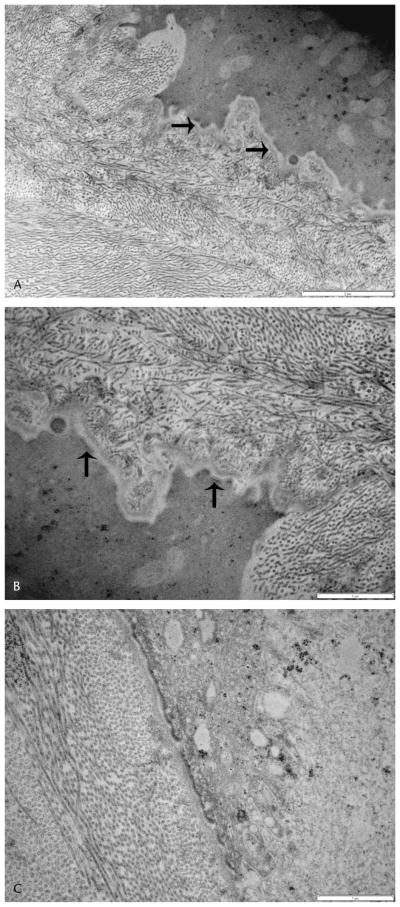
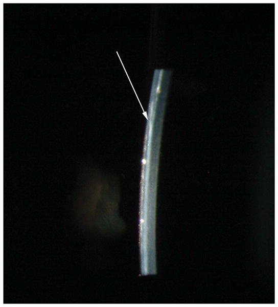
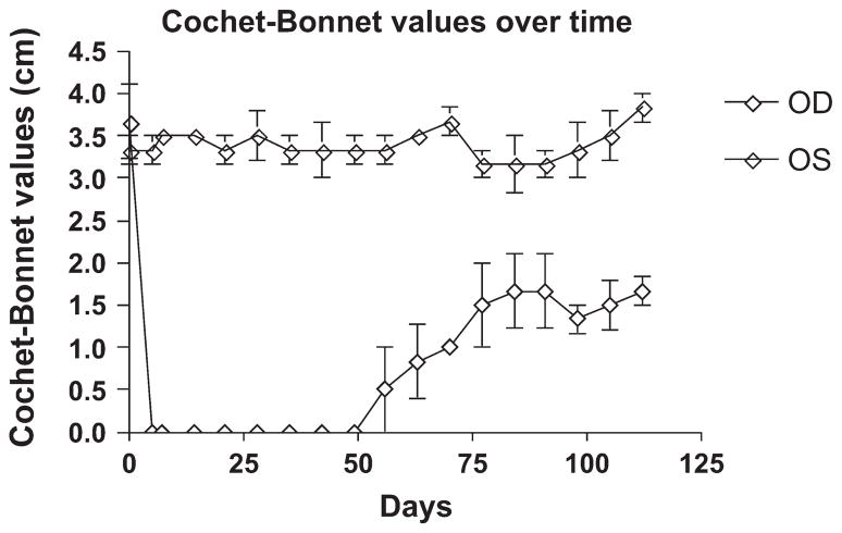
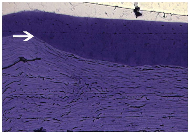
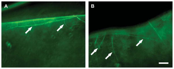
Implants were epithelialized by 7 days. Intraocular pressure was within normal range throughout the study. Aesthesiometry values dropped from an average of 3.67 cm preoperatively to less than 1 mm for all dogs for the first postoperative weeks. By week 16, the average Cochet-Bonnet value was 1.67 cm, demonstrating partial recovery of functional innervation of the implant. No inflammation or rejection of the implant occurred, and minimal haze formation was noted. Light microscopy revealed thickened but normal epithelium over the implant with fibroblast migration into the scaffold. On transmission electron microscopy, the basement membrane was irregular but present and adhesion complexes were noted.
Biosynthetic corneal implantation is well tolerated in dogs, and the collagen-polymer hybrid construct holds promise for clinical application in animals and humans.
评估生物合成角膜植入物在犬体内的整合情况。
三只正常成年实验室比格犬接受了眼科检查,包括裂隙灯生物显微镜检查、间接检眼镜检查、压平眼压测量和 Cochet-Bonnet 触知觉测量。采用改良的表层角膜切除术技术,将戊二醛交联胶原和胶原与聚(N-异丙基丙烯酰胺-co-丙烯酸-co-丙烯酰氧琥珀酰亚胺的共聚物)制成的生物合成角膜植入犬体内。术后第 5 天每天进行眼科检查和触知觉测量,然后每周进行一次,持续 16 周。在第 16 周时,对角膜样本进行组织病理学和透射电子显微镜检查。
植入物在 7 天内被上皮化。整个研究过程中,眼内压均在正常范围内。触知觉值从术前平均 3.67cm 降至所有犬术后前几周的小于 1mm。到第 16 周时,平均 Cochet-Bonnet 值为 1.67cm,表明植入物的功能神经支配部分恢复。植入物无炎症或排斥反应,仅观察到轻微混浊形成。光镜下,可见植入物上有增厚但正常的上皮,成纤维细胞迁移到支架中。在透射电子显微镜下,基底膜不规则但存在,并观察到黏附复合物。
生物合成角膜植入在犬体内耐受良好,胶原-聚合物杂化结构有望在动物和人类的临床应用中得到应用。