Department of Biochemistry and Molecular Biology, University of Calgary, Calgary, Alberta, Canada.
PLoS One. 2010 Jun 7;5(6):e10998. doi: 10.1371/journal.pone.0010998.
Chondrogenesis is the complex process that leads to the establishment of cartilage and bone formation. Due to their ability to differentiate in vitro and mimic development, embryonic stem cells (ESCs) show great potential for investigating developmental processes. In this study, we used chondrogenic differentiation of ESCs as a model to analyze morphogenetic events during chondrogenesis.
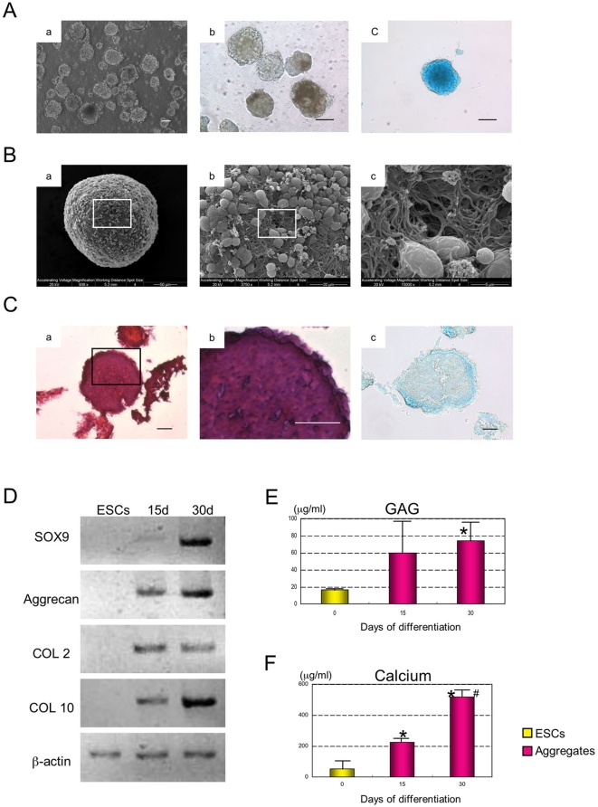
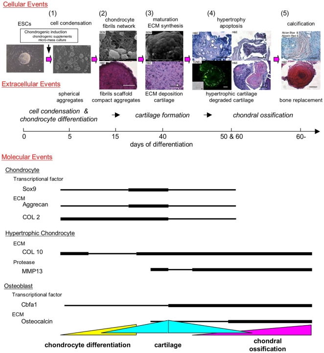
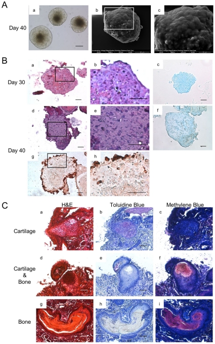
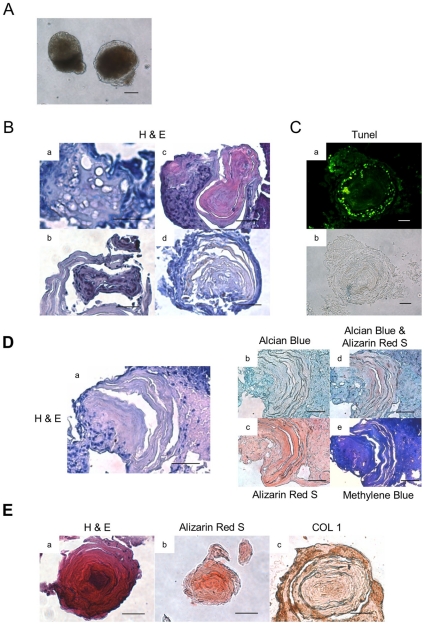
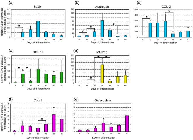
METHODOLOGY/PRINCIPAL FINDINGS: ESCs were differentiated into the chondrocyte lineage, forming small cartilaginous aggregates in suspension. Differentiated ESCs showed that chondrogenesis was typically characterized by five overlapping stages. During the first stage, cell condensation and aggregate formation was observed. The second stage was characterized by differentiation into chondrocytes and fibril scaffold formation within spherical aggregates. Deposition of cartilaginous extracellular matrix and cartilage formation were hallmarks of the third stage. Apoptosis of chondrocytes, hypertrophy and/or degradation of cartilage occurred during the fourth stage. Finally, during the fifth stage, bone replacement with membranous calcified tissues took place.
CONCLUSIONS/SIGNIFICANCE: We demonstrate that ESCs show the chondrogenic differentiation pathway from the pluripotent stem cell to terminal skeletogenesis through these five stages in vitro. During each stage, morphological changes acquired in preceding stages played an important role in further development as a scaffold or template in subsequent stages. The study of chondrogenesis via ESC differentiation may be informative to our further understanding of skeletal growth and regeneration.
软骨发生是导致软骨和骨形成的复杂过程。由于胚胎干细胞(ESCs)具有体外分化的能力并能模拟发育过程,因此它们在研究发育过程方面具有很大的潜力。在这项研究中,我们使用 ESC 的软骨分化作为模型来分析软骨发生过程中的形态发生事件。
方法/主要发现:将 ESCs 分化为成软骨细胞谱系,在悬浮中形成小的软骨聚集物。分化的 ESCs 表明,软骨发生通常具有五个重叠阶段的特征。在第一阶段,观察到细胞凝聚和聚集形成。第二阶段的特征是在球形聚集物中分化为软骨细胞和纤维支架形成。第三阶段的标志是软骨细胞外基质的沉积和软骨形成。第四阶段发生软骨细胞凋亡、肥大和/或软骨降解。最后,在第五阶段,发生膜状钙化组织的骨替代。
结论/意义:我们证明,通过这些体外的五个阶段,ESCs 显示了从多能干细胞到终末成骨的软骨分化途径。在每个阶段,前一阶段获得的形态变化在随后的阶段中作为支架或模板发挥了重要作用,促进了进一步的发育。通过 ESC 分化研究软骨发生可能有助于我们进一步了解骨骼生长和再生。