Owen Dylan M, Oddos Stephane, Kumar Sunil, Davis Daniel M, Neil Mark A A, French Paul M W, Dustin Michael L, Magee Anthony I, Cebecauer Marek
Chemical Biology Centre, Imperial College, London, UK.
Mol Membr Biol. 2010 Aug;27(4-6):178-89. doi: 10.3109/09687688.2010.495353.
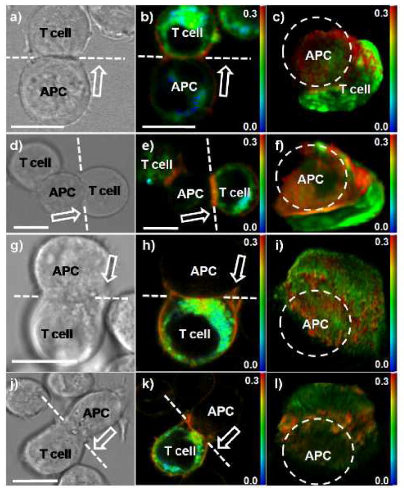
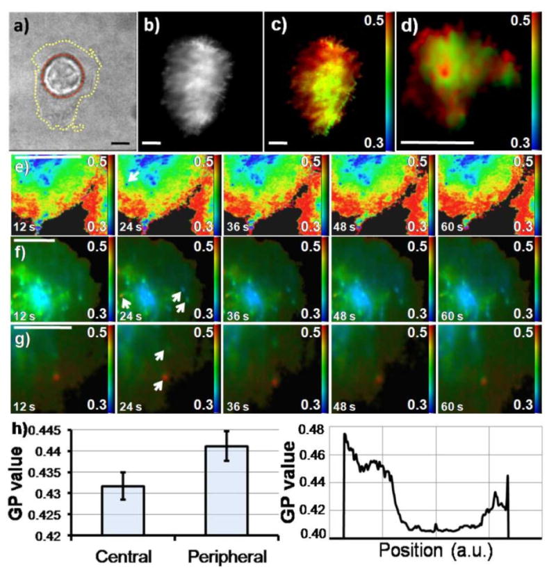
Cholesterol- and glycosphingolipid-enriched membrane lipid microdomains, frequently called lipid rafts, are thought to play an important role in the spatial and temporal organization of immunological synapses. Higher ordering of lipid acyl chains was suggested for these entities and imaging of membrane order in living cells during activation can therefore help to understand the mechanisms responsible for the supramolecular organization of molecules involved in the activation of T cells. Here, we employ the phase-sensitive membrane dye di-4-ANEPPDHQ together with a variety of spectrally-resolved microscopy techniques, including 2-channel ratiometric TIRF microscopy and fluorescence lifetime imaging, to characterize membrane order at the T cell immunological synapse at high spatial and temporal resolution in live cells at physiological temperature. We find that higher membrane order resides at the immunological synapse periphery where proximal signalling through the immunoreceptors and accessory proteins in microclusters has previously been shown to take place. The observed spatial patterning of membrane order in the immunological synapse depends on active receptor signalling.
富含胆固醇和糖鞘脂的膜脂微区,通常被称为脂筏,被认为在免疫突触的时空组织中发挥重要作用。这些实体的脂质酰基链被认为具有更高的有序性,因此在激活过程中对活细胞中的膜有序性进行成像有助于理解负责T细胞激活中分子超分子组织的机制。在这里,我们使用相敏膜染料di-4-ANEPPDHQ以及各种光谱分辨显微镜技术,包括双通道比率TIRF显微镜和荧光寿命成像,以在生理温度下的活细胞中以高时空分辨率表征T细胞免疫突触处的膜有序性。我们发现更高的膜有序性存在于免疫突触周边,此前已表明通过微簇中的免疫受体和辅助蛋白进行的近端信号传导在此处发生。免疫突触中观察到的膜有序性空间模式取决于活性受体信号传导。