• 文献检索
  • 文档翻译
  • 深度研究
  • 学术资讯
  • Suppr Zotero 插件Zotero 插件
  • 邀请有礼
  • 套餐&价格
  • 历史记录
应用&插件
Suppr Zotero 插件Zotero 插件浏览器插件Mac 客户端Windows 客户端微信小程序
定价
高级版会员购买积分包购买API积分包
服务
文献检索文档翻译深度研究API 文档MCP 服务
关于我们
关于 Suppr公司介绍联系我们用户协议隐私条款
关注我们

Suppr 超能文献

核心技术专利:CN118964589B侵权必究
粤ICP备2023148730 号-1Suppr @ 2026

文献检索

告别复杂PubMed语法,用中文像聊天一样搜索,搜遍4000万医学文献。AI智能推荐,让科研检索更轻松。

立即免费搜索

文件翻译

保留排版,准确专业,支持PDF/Word/PPT等文件格式,支持 12+语言互译。

免费翻译文档

深度研究

AI帮你快速写综述,25分钟生成高质量综述,智能提取关键信息,辅助科研写作。

立即免费体验

动脉弹性成像:有限元分析模型与高分辨率超声斑点追踪的比较

Arterial elasticity imaging: comparison of finite-element analysis models with high-resolution ultrasound speckle tracking.

作者信息

Park Dae Woo, Richards Michael S, Rubin Jonathan M, Hamilton James, Kruger Grant H, Weitzel William F

机构信息

Department of Internal Medicine, University of Michigan, Ann Arbor, Michigan, USA.

出版信息

Cardiovasc Ultrasound. 2010 Jun 18;8:22. doi: 10.1186/1476-7120-8-22.

DOI:10.1186/1476-7120-8-22
PMID:20565833
原文链接:https://pmc.ncbi.nlm.nih.gov/articles/PMC2908573/
Abstract

BACKGROUND

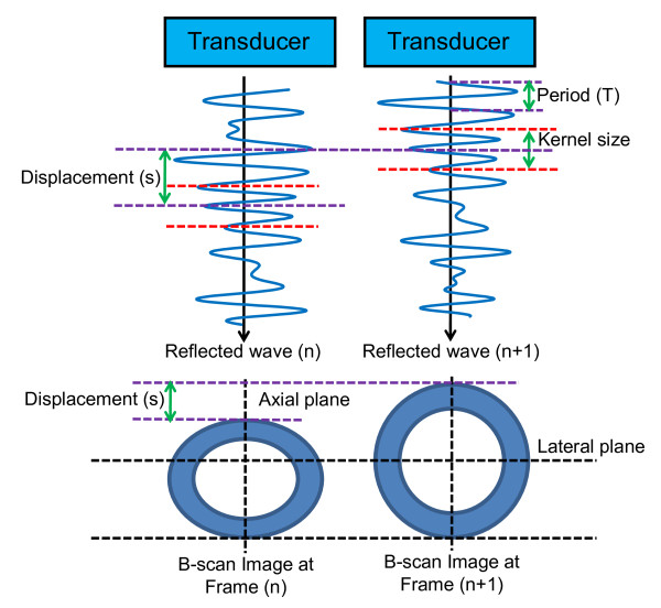
The nonlinear mechanical properties of internal organs and tissues may be measured with unparalleled precision using ultrasound imaging with phase-sensitive speckle tracking. The many potential applications of this important noninvasive diagnostic approach include measurement of arterial stiffness, which is associated with numerous major disease processes. The accuracy of previous ultrasound measurements of arterial stiffness and vascular elasticity has been limited by the relatively low strain of nonlinear structures under normal physiologic pressure and the measurement assumption that the effect of the surrounding tissue modulus might be ignored in both physiologic and pressure equalized conditions.

METHODS

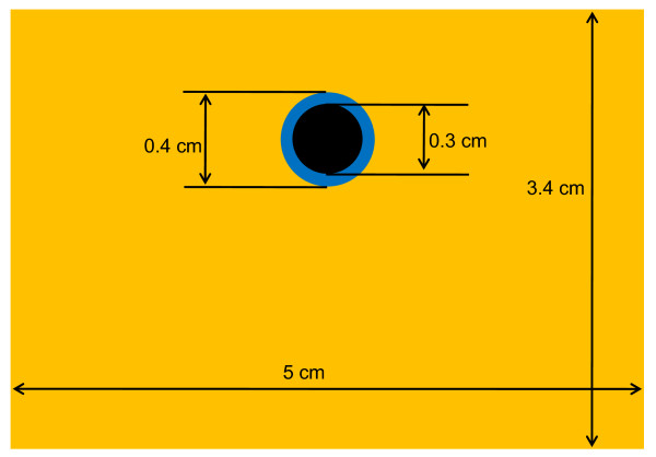
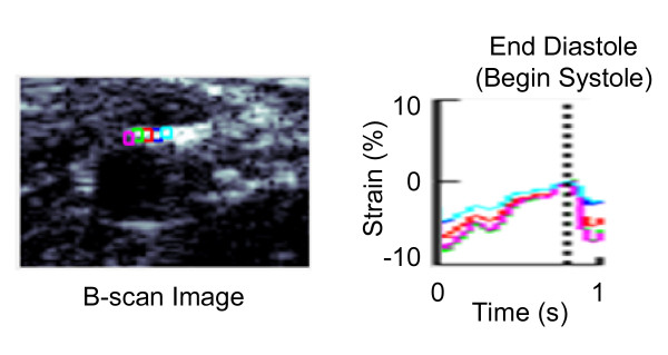
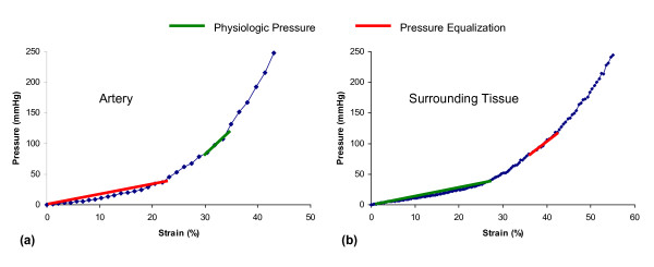
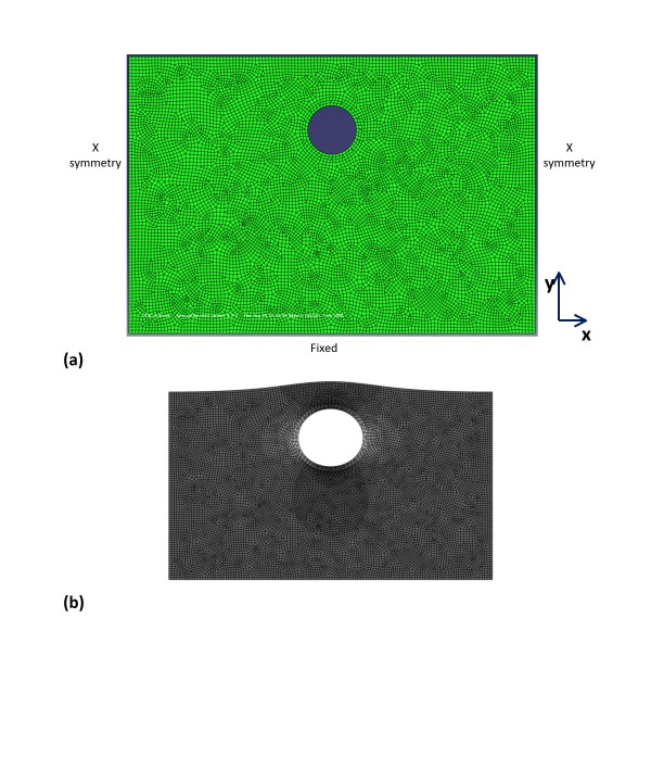
This study performed high-resolution ultrasound imaging of the brachial artery in a healthy adult subject under normal physiologic pressure and the use of external pressure (pressure equalization) to increase strain. These ultrasound results were compared to measurements of arterial strain as determined by finite-element analysis models with and without a surrounding tissue, which was represented by homogenous material with fixed elastic modulus.

RESULTS

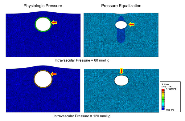
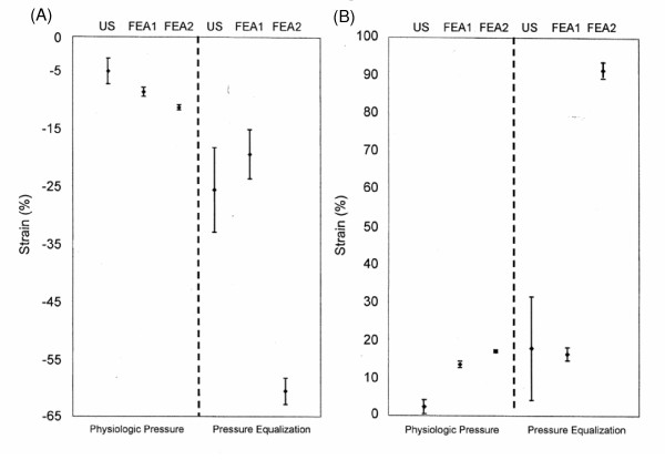
Use of the pressure equalization technique during imaging resulted in average strain values of 26% and 18% at the top and sides, respectively, compared to 5% and 2%, at the top and sides, respectively, under physiologic pressure. In the artery model that included surrounding tissue, strain was 19% and 16% under pressure equalization versus 9% and 13% at the top and sides, respectively, under physiologic pressure. The model without surrounding tissue had slightly higher levels of strain under physiologic pressure compared to the other model, but the resulting strain values under pressure equalization were > 60% and did not correspond to experimental values.

CONCLUSIONS

Since pressure equalization may increase the dynamic range of strain imaging, the effect of the surrounding tissue on strain should be incorporated into models of arterial strain, particularly when the pressure equalization technique is used.

摘要

背景

使用具有相位敏感散斑跟踪的超声成像可以以前所未有的精度测量内部器官和组织的非线性力学特性。这种重要的非侵入性诊断方法有许多潜在应用,包括测量与众多主要疾病过程相关的动脉僵硬度。以往超声测量动脉僵硬度和血管弹性的准确性受到正常生理压力下非线性结构相对较低应变以及测量假设的限制,该假设认为在生理和压力均衡条件下周围组织模量的影响可能被忽略。

方法

本研究对一名健康成年受试者的肱动脉在正常生理压力下进行了高分辨率超声成像,并使用外部压力(压力均衡)来增加应变。将这些超声结果与通过有限元分析模型确定的动脉应变测量值进行比较,该模型有或没有周围组织,周围组织由具有固定弹性模量的均匀材料表示。

结果

成像过程中使用压力均衡技术时,顶部和侧面的平均应变值分别为26%和18%,而在生理压力下,顶部和侧面的平均应变值分别为5%和2%。在包含周围组织的动脉模型中,压力均衡下的应变分别为19%和16%,而在生理压力下,顶部和侧面的应变分别为9%和13%。与另一个模型相比,没有周围组织的模型在生理压力下的应变水平略高,但在压力均衡下得到的应变值>60%,且与实验值不相符。

结论

由于压力均衡可能增加应变成像的动态范围,因此在动脉应变模型中应纳入周围组织对应变的影响,特别是在使用压力均衡技术时。

https://cdn.ncbi.nlm.nih.gov/pmc/blobs/f8c5/2908573/464b233ce6cd/1476-7120-8-22-7.jpg
https://cdn.ncbi.nlm.nih.gov/pmc/blobs/f8c5/2908573/2d24d31945a4/1476-7120-8-22-1.jpg
https://cdn.ncbi.nlm.nih.gov/pmc/blobs/f8c5/2908573/28a14e445630/1476-7120-8-22-2.jpg
https://cdn.ncbi.nlm.nih.gov/pmc/blobs/f8c5/2908573/1d83e7593973/1476-7120-8-22-3.jpg
https://cdn.ncbi.nlm.nih.gov/pmc/blobs/f8c5/2908573/2fbee5408aed/1476-7120-8-22-4.jpg
https://cdn.ncbi.nlm.nih.gov/pmc/blobs/f8c5/2908573/19f78306f71b/1476-7120-8-22-5.jpg
https://cdn.ncbi.nlm.nih.gov/pmc/blobs/f8c5/2908573/50a38fcf1fe8/1476-7120-8-22-6.jpg
https://cdn.ncbi.nlm.nih.gov/pmc/blobs/f8c5/2908573/464b233ce6cd/1476-7120-8-22-7.jpg

相似文献

1
Arterial elasticity imaging: comparison of finite-element analysis models with high-resolution ultrasound speckle tracking.动脉弹性成像:有限元分析模型与高分辨率超声斑点追踪的比较
Cardiovasc Ultrasound. 2010 Jun 18;8:22. doi: 10.1186/1476-7120-8-22.
2
Vascular intramural strain imaging using arterial pressure equalization.利用动脉压力均衡进行血管壁内应变成像。
Ultrasound Med Biol. 2004 Jun;30(6):761-71. doi: 10.1016/j.ultrasmedbio.2004.04.003.
3
In vivo vascular wall shear rate and circumferential strain of renal disease patients.肾病患者血管壁切变率和周向应变的体内测量。
Ultrasound Med Biol. 2013 Feb;39(2):241-52. doi: 10.1016/j.ultrasmedbio.2012.08.027. Epub 2012 Dec 1.
4
Ultrasound measurement of brachial artery elasticity prior to hemodialysis access placement: a pilot study.超声测量肱动脉弹性在血液透析通路建立前:一项初步研究。
J Ultrasound Med. 2012 Oct;31(10):1581-8. doi: 10.7863/jum.2012.31.10.1581.
5
Direct effects of smooth muscle relaxation and contraction on in vivo human brachial artery elastic properties.平滑肌舒张和收缩对体内人体肱动脉弹性特性的直接影响。
Circ Res. 1995 Nov;77(5):1008-16. doi: 10.1161/01.res.77.5.1008.
6
A new nonlinear parameter in the developed strain-to-applied strain of the soft tissues and its application in ultrasound elasticity imaging.一种软组织应变成形中的新非线性参数及其在超声弹性成像中的应用。
Ultrasound Med Biol. 2012 Mar;38(3):511-23. doi: 10.1016/j.ultrasmedbio.2011.12.009. Epub 2012 Jan 21.
7
Comparing Carotid and Brachial Artery Stiffness: A First Step Toward Mechanical Mapping of the Arterial Tree.比较颈动脉和肱动脉僵硬度:迈向动脉树力学图谱绘制的第一步。
Ultrasound Med Biol. 2015 Jul;41(7):1808-13. doi: 10.1016/j.ultrasmedbio.2015.02.013. Epub 2015 Apr 1.
8
Contribution of collagen, elastin, and smooth muscle to in vivo human brachial artery wall stress and elastic modulus.胶原蛋白、弹性蛋白和平滑肌对人体肱动脉体内壁应力和弹性模量的贡献。
Circulation. 1996 Dec 15;94(12):3263-70. doi: 10.1161/01.cir.94.12.3263.
9
In vivo human brachial artery elastic mechanics: effects of smooth muscle relaxation.体内人体肱动脉弹性力学:平滑肌舒张的影响
Circulation. 1999 Jul 6;100(1):41-7. doi: 10.1161/01.cir.100.1.41.
10
High-resolution ultrasound speckle tracking may detect vascular mechanical wall changes in peripheral artery bypass vein grafts.高分辨率超声斑点追踪技术可能检测出外周动脉搭桥静脉移植物中的血管壁机械变化。
Ann Vasc Surg. 2009 Mar;23(2):201-6. doi: 10.1016/j.avsg.2008.08.031. Epub 2008 Oct 29.

引用本文的文献

1
Quantification of the heterogeneous effect of static and dynamic perivascular structures on patient-specific local aortic wall mechanics using inverse finite element modeling and DENSE MRI.利用逆有限元建模和 DENSE MRI 定量评估静态和动态血管周围结构对患者特定局部主动脉壁力学的异质影响。
J Biomech. 2022 Jun;138:111119. doi: 10.1016/j.jbiomech.2022.111119. Epub 2022 May 5.
2
Plaque hemorrhage in carotid artery disease: pathogenesis, clinical and biomechanical considerations.颈动脉疾病中的斑块出血:发病机制、临床和生物力学考虑。
J Biomech. 2014 Mar 3;47(4):847-58. doi: 10.1016/j.jbiomech.2014.01.013. Epub 2014 Jan 13.
3

本文引用的文献

1
High-resolution ultrasound elasticity imaging to evaluate dialysis fistula stenosis.高分辨率超声弹性成像评估透析瘘管狭窄
Semin Dial. 2009 Jan-Feb;22(1):84-9. doi: 10.1111/j.1525-139X.2008.00502.x. Epub 2008 Sep 24.
2
In vivo vibroacoustography of large peripheral arteries.大型外周动脉的体内振动声学成像
Invest Radiol. 2008 Apr;43(4):243-52. doi: 10.1097/RLI.0b013e31816085fc.
3
Speckle tracking methods for ultrasonic elasticity imaging using short-time correlation.基于短时相关性的超声弹性成像散斑追踪方法。
Non-invasive speed of sound measurement in cartilage by use of combined magnetic resonance imaging and ultrasound: an initial study.
利用磁共振成像和超声联合技术对软骨进行非侵入性声速测量:一项初步研究。
Radiol Phys Technol. 2013 Jul;6(2):480-5. doi: 10.1007/s12194-013-0223-4. Epub 2013 Jun 1.
4
In vivo vascular wall shear rate and circumferential strain of renal disease patients.肾病患者血管壁切变率和周向应变的体内测量。
Ultrasound Med Biol. 2013 Feb;39(2):241-52. doi: 10.1016/j.ultrasmedbio.2012.08.027. Epub 2012 Dec 1.
5
Ultrasound measurement of brachial artery elasticity prior to hemodialysis access placement: a pilot study.超声测量肱动脉弹性在血液透析通路建立前:一项初步研究。
J Ultrasound Med. 2012 Oct;31(10):1581-8. doi: 10.7863/jum.2012.31.10.1581.
IEEE Trans Ultrason Ferroelectr Freq Control. 1999;46(1):82-96. doi: 10.1109/58.741427.
4
Noninvasive generation and measurement of propagating waves in arterial walls.动脉壁中传播波的无创产生与测量。
J Acoust Soc Am. 2006 Feb;119(2):1238-43. doi: 10.1121/1.2159294.
5
Evaluation of carotid-femoral pulse wave velocity: influence of timing algorithm and heart rate.颈动脉-股动脉脉搏波速度的评估:定时算法和心率的影响
Hypertension. 2005 Feb;45(2):222-6. doi: 10.1161/01.HYP.0000154229.97341.d2. Epub 2005 Jan 10.
6
Renal advances in ultrasound elasticity imaging: measuring the compliance of arteries and kidneys in end-stage renal disease.超声弹性成像在肾脏领域的进展:测量终末期肾病患者的动脉和肾脏顺应性
Blood Purif. 2005;23(1):10-7. doi: 10.1159/000082005.
7
Vascular intramural strain imaging using arterial pressure equalization.利用动脉压力均衡进行血管壁内应变成像。
Ultrasound Med Biol. 2004 Jun;30(6):761-71. doi: 10.1016/j.ultrasmedbio.2004.04.003.
8
Strain imaging of internal deformation.内部变形的应变成像
Ultrasound Med Biol. 2002 Nov-Dec;28(11-12):1475-84. doi: 10.1016/s0301-5629(02)00645-2.
9
Methods and devices for measuring arterial compliance in humans.测量人体动脉顺应性的方法和装置。
Am J Hypertens. 2002 Aug;15(8):743-53. doi: 10.1016/s0895-7061(02)02962-x.
10
Arterial pulse wave velocity with tissue Doppler imaging.组织多普勒成像测量动脉脉搏波速度
Ultrasound Med Biol. 2002 May;28(5):571-80. doi: 10.1016/s0301-5629(02)00495-7.