• 文献检索
  • 文档翻译
  • 深度研究
  • 学术资讯
  • Suppr Zotero 插件Zotero 插件
  • 邀请有礼
  • 套餐&价格
  • 历史记录
应用&插件
Suppr Zotero 插件Zotero 插件浏览器插件Mac 客户端Windows 客户端微信小程序
定价
高级版会员购买积分包购买API积分包
服务
文献检索文档翻译深度研究API 文档MCP 服务
关于我们
关于 Suppr公司介绍联系我们用户协议隐私条款
关注我们

Suppr 超能文献

核心技术专利:CN118964589B侵权必究
粤ICP备2023148730 号-1Suppr @ 2026

文献检索

告别复杂PubMed语法,用中文像聊天一样搜索,搜遍4000万医学文献。AI智能推荐,让科研检索更轻松。

立即免费搜索

文件翻译

保留排版,准确专业,支持PDF/Word/PPT等文件格式,支持 12+语言互译。

免费翻译文档

深度研究

AI帮你快速写综述,25分钟生成高质量综述,智能提取关键信息,辅助科研写作。

立即免费体验

鸭肠炎病毒 pUL38 蛋白的表达与细胞内定位。

Expression and intracellular localization of duck enteritis virus pUL38 protein.

机构信息

Avian Diseases Research Center, College of Veterinary Medicine of Sichuan Agricultural University, Yaan, Sichuan, China.

出版信息

Virol J. 2010 Jul 17;7:162. doi: 10.1186/1743-422X-7-162.

DOI:10.1186/1743-422X-7-162
PMID:20637115
原文链接:https://pmc.ncbi.nlm.nih.gov/articles/PMC2918563/
Abstract

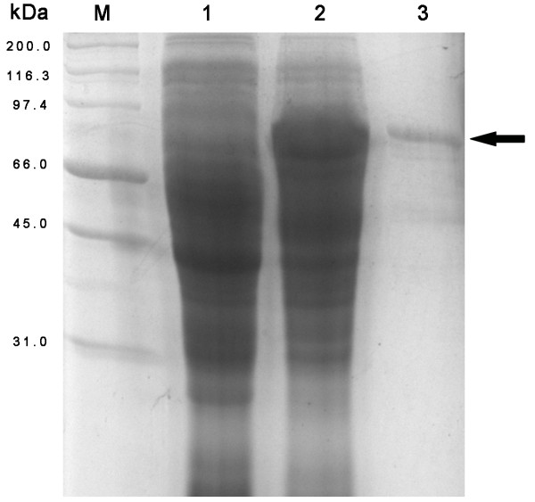
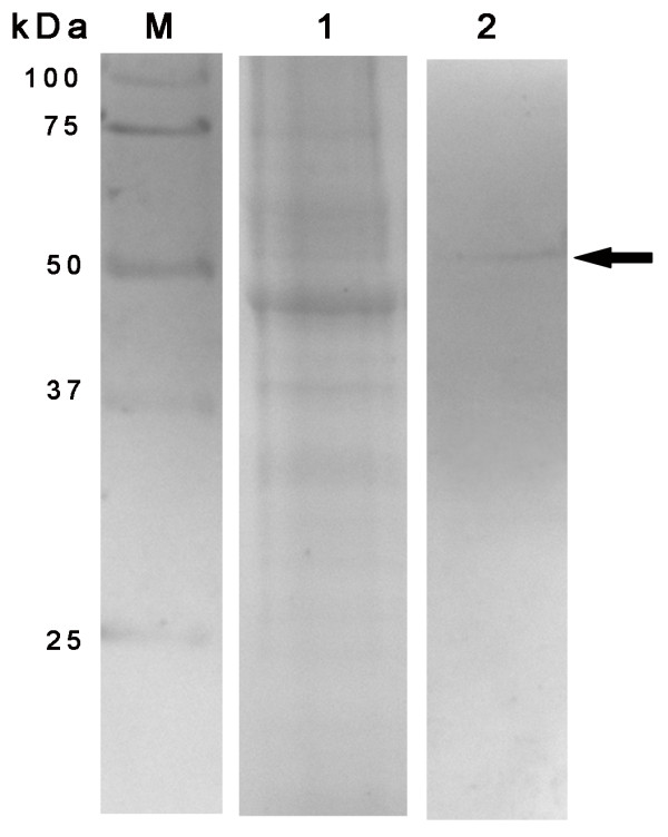
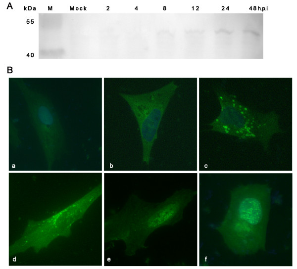
Knowledge of the intracellular location of a protein can provide useful insights into its function. Bioinformatic studies have predicted that the DEV pUL38 mainly targets the cytoplasm and nucleus. In this study, we obtained anti-pUL38 polyclonal sera. These antibodies were functional in western blotting and immunofluorescence in DEV-infected duck embryo fibroblasts (DEFs). pUL38 was expressed as a 51-kDa protein from 8 h post-infection onward, initially showing a diffuse distribution throughout the cytoplasm, and later in the nucleus. Furthermore, pUL38 was found in purified virus. These results provide the first evidence of the kinetics of expression and intracellular localization of DEV pUL38.

摘要

蛋白质的细胞内位置的知识可以为其功能提供有用的见解。生物信息学研究预测,DEV pUL38 主要靶向细胞质和细胞核。在这项研究中,我们获得了抗 pUL38 多克隆血清。这些抗体在 DEV 感染的鸭胚成纤维细胞(DEFs)中的 western blot 和免疫荧光中具有功能。pUL38 在感染后 8 小时开始作为 51kDa 的蛋白表达,最初在细胞质中呈弥散分布,随后在细胞核中分布。此外,pUL38 存在于纯化的病毒中。这些结果首次提供了 DEV pUL38 的表达和细胞内定位动力学的证据。

https://cdn.ncbi.nlm.nih.gov/pmc/blobs/b3c8/2918563/e81f6bc0e88b/1743-422X-7-162-3.jpg
https://cdn.ncbi.nlm.nih.gov/pmc/blobs/b3c8/2918563/4e6121314f41/1743-422X-7-162-1.jpg
https://cdn.ncbi.nlm.nih.gov/pmc/blobs/b3c8/2918563/afa31f078e93/1743-422X-7-162-2.jpg
https://cdn.ncbi.nlm.nih.gov/pmc/blobs/b3c8/2918563/e81f6bc0e88b/1743-422X-7-162-3.jpg
https://cdn.ncbi.nlm.nih.gov/pmc/blobs/b3c8/2918563/4e6121314f41/1743-422X-7-162-1.jpg
https://cdn.ncbi.nlm.nih.gov/pmc/blobs/b3c8/2918563/afa31f078e93/1743-422X-7-162-2.jpg
https://cdn.ncbi.nlm.nih.gov/pmc/blobs/b3c8/2918563/e81f6bc0e88b/1743-422X-7-162-3.jpg

相似文献

1
Expression and intracellular localization of duck enteritis virus pUL38 protein.鸭肠炎病毒 pUL38 蛋白的表达与细胞内定位。
Virol J. 2010 Jul 17;7:162. doi: 10.1186/1743-422X-7-162.
2
Replication kinetics of duck enteritis virus UL16 gene in vitro.鸭肠炎病毒 UL16 基因在体外的复制动力学。
Virol J. 2012 Nov 21;9:281. doi: 10.1186/1743-422X-9-281.
3
Molecular characterization of the duck enteritis virus US10 protein.鸭肠炎病毒 US10 蛋白的分子特征。
Virol J. 2017 Sep 20;14(1):183. doi: 10.1186/s12985-017-0841-2.
4
Complete genomic sequence of Chinese virulent duck enteritis virus.中国强毒鸭肠炎病毒的全基因组序列。
J Virol. 2012 May;86(10):5965. doi: 10.1128/JVI.00529-12.
5
Duck enteritis virus UL21 is a late gene encoding a protein that interacts with pUL16.鸭肠炎病毒 UL21 是一个晚期基因,编码一种与 pUL16 相互作用的蛋白。
BMC Vet Res. 2020 Jan 8;16(1):8. doi: 10.1186/s12917-019-2228-7.
6
Expression and characterization of duck enteritis virus gI gene.鸭肠炎病毒 gI 基因的表达与鉴定。
Virol J. 2011 May 19;8:241. doi: 10.1186/1743-422X-8-241.
7
Duck enteritis virus pUL47, as a late structural protein localized in the nucleus, mainly depends on residues 40 to 50 and 768 to 777 and inhibits IFN-β signalling by interacting with STAT1.鸭肠炎病毒 pUL47 作为一种晚期核定位结构蛋白,主要依赖于残基 40 到 50 和 768 到 777 并通过与 STAT1 相互作用抑制 IFN-β 信号转导。
Vet Res. 2020 Nov 11;51(1):135. doi: 10.1186/s13567-020-00859-w.
8
Prokaryotic expression of the truncated duck enteritis virus UL27 gene and characteristics of UL27 gene and its truncated product.鸭肠炎病毒UL27基因截短体的原核表达及UL27基因及其截短产物的特性
Acta Virol. 2012;56(4):323-8. doi: 10.4149/av_2012_04_323.
9
Adaptation and growth kinetics study of an Indian isolate of virulent duck enteritis virus in Vero cells.一株强毒鸭肠炎病毒印度分离株在Vero细胞中的适应性及生长动力学研究
Microb Pathog. 2015 Jan;78:14-9. doi: 10.1016/j.micpath.2014.11.008. Epub 2014 Nov 13.
10
Complete genome sequence of an isolate of duck enteritis virus from China.中国鸭肠炎病毒分离株的全基因组序列。
Arch Virol. 2020 Jul;165(7):1687-1689. doi: 10.1007/s00705-020-04594-7. Epub 2020 May 8.

引用本文的文献

1
Pseudorabies virus UL38 attenuates the cGAS-STING signaling pathway by recruiting Tollip to promote STING for autophagy degradation.伪狂犬病毒 UL38 通过招募 Tollip 来抑制 cGAS-STING 信号通路,从而促进 STING 发生自噬降解。
Virol J. 2024 May 8;21(1):107. doi: 10.1186/s12985-024-02379-x.
2
Duck enteritis virus pUL47, as a late structural protein localized in the nucleus, mainly depends on residues 40 to 50 and 768 to 777 and inhibits IFN-β signalling by interacting with STAT1.鸭肠炎病毒 pUL47 作为一种晚期核定位结构蛋白,主要依赖于残基 40 到 50 和 768 到 777 并通过与 STAT1 相互作用抑制 IFN-β 信号转导。
Vet Res. 2020 Nov 11;51(1):135. doi: 10.1186/s13567-020-00859-w.
3

本文引用的文献

1
Characterization of duck enteritis virus US6, US7 and US8 gene.鸭肠炎病毒 US6、US7 和 US8 基因的特征。
Intervirology. 2010;53(3):141-5. doi: 10.1159/000274974. Epub 2010 Jan 13.
2
Identification and characterization of a duck enteritis virus US3-like gene.鸭肠炎病毒US3样基因的鉴定与特性分析
Avian Dis. 2009 Sep;53(3):363-9. doi: 10.1637/8643-020409-Reg.1.
3
Cloning, expression, purification and characterization of UL24 partial protein of duck enteritis virus.鸭肠炎病毒 UL24 部分蛋白的克隆、表达、纯化及鉴定
Duck Enteritis Virus VP16 Antagonizes IFN--Mediated Antiviral Innate Immunity.
鸭肠炎病毒 VP16 拮抗 IFN--介导的抗病毒先天免疫。
J Immunol Res. 2020 May 15;2020:9630452. doi: 10.1155/2020/9630452. eCollection 2020.
4
US10 Protein Is Crucial but not Indispensable for Duck Enteritis Virus Infection in Vitro.US10 蛋白对鸭肠炎病毒体外感染至关重要但非必需。
Sci Rep. 2018 Nov 7;8(1):16510. doi: 10.1038/s41598-018-34503-7.
5
Molecular characterization of duck enteritis virus UL41 protein.鸭肠炎病毒 UL41 蛋白的分子特征。
Virol J. 2018 Jan 15;15(1):12. doi: 10.1186/s12985-018-0928-4.
6
Duck enteritis virus (DEV) UL54 protein, a novel partner, interacts with DEV UL24 protein.鸭肠炎病毒(DEV)UL54蛋白是一种新型伴侣蛋白,可与DEV UL24蛋白相互作用。
Virol J. 2017 Aug 29;14(1):166. doi: 10.1186/s12985-017-0830-5.
7
Replication kinetics of duck enteritis virus UL16 gene in vitro.鸭肠炎病毒 UL16 基因在体外的复制动力学。
Virol J. 2012 Nov 21;9:281. doi: 10.1186/1743-422X-9-281.
8
Comparative genomic analysis of duck enteritis virus strains.鸭肠炎病毒株的比较基因组分析。
J Virol. 2012 Dec;86(24):13841-2. doi: 10.1128/JVI.01517-12.
9
Expression and characterization of UL16 gene from duck enteritis virus.鸭肠炎病毒 UL16 基因的表达与鉴定。
Virol J. 2011 Aug 24;8:413. doi: 10.1186/1743-422X-8-413.
10
Characterization of the duck enteritis virus UL55 protein.鸭肠炎病毒 UL55 蛋白的特性分析。
Virol J. 2011 May 24;8:256. doi: 10.1186/1743-422X-8-256.
Intervirology. 2009;52(6):326-34. doi: 10.1159/000242354. Epub 2009 Sep 29.
4
Herpesvirus assembly: an update.疱疹病毒组装:最新进展。
Virus Res. 2009 Aug;143(2):222-34. doi: 10.1016/j.virusres.2009.03.018. Epub 2009 Apr 7.
5
Characterization of subcellular localization of duck enteritis virus UL51 protein.鸭肠炎病毒UL51蛋白的亚细胞定位特征
Virol J. 2009 Jul 3;6:92. doi: 10.1186/1743-422X-6-92.
6
Identification and characterization of the duck enteritis virus UL51 gene.鸭肠炎病毒UL51基因的鉴定与特性分析
Arch Virol. 2009;154(7):1061-9. doi: 10.1007/s00705-009-0407-8. Epub 2009 Jun 11.
7
His6-tagged UL35 protein of duck plague virus: expression, purification, and production of polyclonal antibody.鸭瘟病毒His6标签化的UL35蛋白:表达、纯化及多克隆抗体的制备
Intervirology. 2009;52(3):141-51. doi: 10.1159/000221833. Epub 2009 May 29.
8
Molecular cloning and characterization of the UL31 gene from duck enteritis virus.鸭肠炎病毒 UL31 基因的克隆与特性分析。
Mol Biol Rep. 2010 Mar;37(3):1495-503. doi: 10.1007/s11033-009-9546-y. Epub 2009 May 23.
9
Anatid herpesvirus 1 CH virulent strain induces syncytium and apoptosis in duck embryo fibroblast cultures.鸭疱疹病毒1型CH强毒株在鸭胚成纤维细胞培养物中诱导形成多核巨细胞和凋亡。
Vet Microbiol. 2009 Sep 18;138(3-4):258-65. doi: 10.1016/j.vetmic.2009.04.006. Epub 2009 Apr 10.
10
Expression and characterization of the UL31 protein from duck enteritis virus.鸭肠炎病毒UL31蛋白的表达与鉴定
Virol J. 2009 Feb 10;6:19. doi: 10.1186/1743-422X-6-19.