Tanaka Sai-ichi, Sumioka Takayoshi, Fujita Norihito, Kitano Ai, Okada Yuka, Yamanaka Osamu, Flanders Kathleen C, Miyajima Masayasu, Saika Shizuya
Department of Ophthalmology, Wakayama Medical University, Wakayama, Japan.
Mol Vis. 2010 Jul 1;16:1194-205.
To investigate the role of tenascin-C in epithelial-mesenchymal transition (EMT) of the lens epithelium during wound healing in mice. Tenascin-C is a component of the extracellular matrix in patients having post-operative capsular opacification.
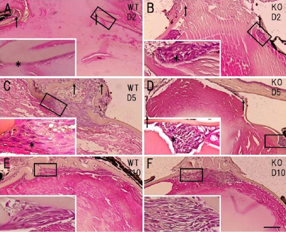
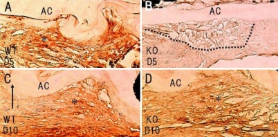
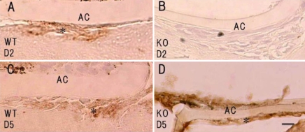
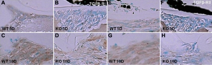
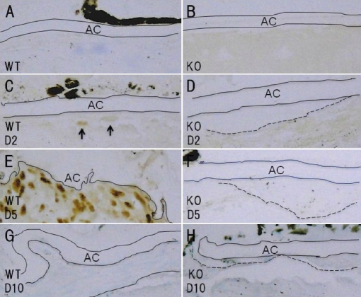
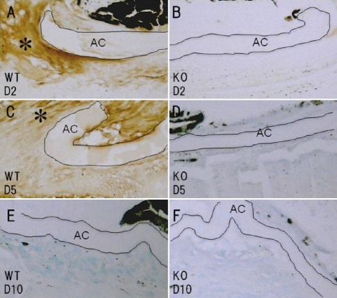
The crystalline lens was injured by needle puncture in tenascin-C null (KO, n=56) and wild-type (WT, n=56) mice in a C57BL/6 background. The animals were killed at day 2, 5, or 10 post-injury. Immunohistochemistry was employed to detect alpha-smooth muscle actin (alphaSMA), a marker of EMT, collagen type I, transforming growth factor beta1 (TGFbeta1), phospho-Smad2, phospho-adducin, and phospho-myosin light chain 9 (MLC9). The expression levels of phospho-adducin and phospho-MLC9 were used as markers for the activation of protein kinase C and Rho kinase, respectively.
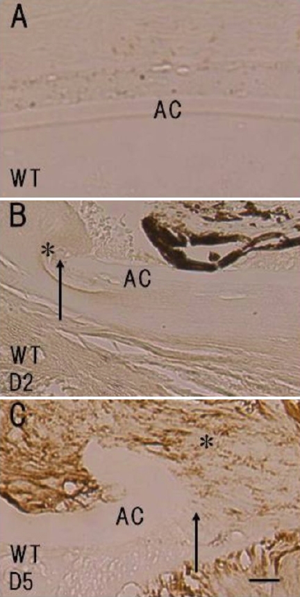
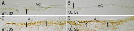
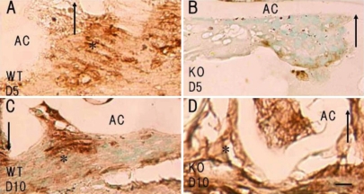
The expression of tenascin-C was upregulated in WT lens epithelial cells adjacent to the capsular break at day 5. The results showed that injury-induced EMT of the mouse lens epithelium, as evaluated by histology and the expression patterns of alphaSMA and fibronectin, was attenuated in the absence of tenascin-C. Upregulation of TGFbeta1 expression in the epithelium was also inhibited, and loss of tenascin-C attenuated the phosphorylation of Smad2 and adducin in epithelial cells adjacent to the capsular break. The expression of phospho-adducin was suppressed, while the expression level of phospho-MLC9 was unchanged, in the healing epithelium in the absence of tenascin C.
Tenascin-C is required for injury-induced EMT in the mouse lens epithelium. The mechanism behind this might involve impaired activation of cytoplasmic signaling cascades; i.e., TGFbeta/Smad and protein kinase C-adducing signaling, in the absence of tenascin-C.
研究肌腱蛋白-C在小鼠伤口愈合过程中晶状体上皮细胞上皮-间质转化(EMT)中的作用。肌腱蛋白-C是术后囊膜混浊患者细胞外基质的一个组成部分。
在C57BL/6背景的肌腱蛋白-C基因敲除(KO,n = 56)和野生型(WT,n = 56)小鼠中,用针刺损伤晶状体。在损伤后第2、5或10天处死动物。采用免疫组织化学法检测α-平滑肌肌动蛋白(αSMA)(EMT的标志物)、I型胶原、转化生长因子β1(TGFβ1)、磷酸化Smad2、磷酸化内收蛋白和磷酸化肌球蛋白轻链9(MLC9)。磷酸化内收蛋白和磷酸化MLC9的表达水平分别用作蛋白激酶C和Rho激酶激活的标志物。
在第5天,WT晶状体上皮细胞中靠近囊膜破裂处的肌腱蛋白-C表达上调。结果表明,通过组织学以及αSMA和纤连蛋白的表达模式评估,在缺乏肌腱蛋白-C的情况下,损伤诱导的小鼠晶状体上皮细胞EMT减弱。上皮细胞中TGFβ1表达的上调也受到抑制,并且肌腱蛋白-C的缺失减弱了靠近囊膜破裂处上皮细胞中Smad2和内收蛋白的磷酸化。在缺乏肌腱蛋白-C的情况下,愈合上皮中磷酸化内收蛋白的表达受到抑制,而磷酸化MLC9的表达水平未改变。
肌腱蛋白-C是小鼠晶状体上皮细胞损伤诱导的EMT所必需的。其背后的机制可能涉及在缺乏肌腱蛋白-C的情况下,细胞质信号级联反应(即TGFβ/Smad和蛋白激酶C-内收蛋白信号传导)的激活受损。