MRC Human Reproductive Sciences Unit, Centre for Reproductive Biology, The Queen's Medical Research Institute, University of Edinburgh, 47 Little France Crescent, Edinburgh EH16 4TJ, UK.
Hum Reprod. 2010 Oct;25(10):2405-14. doi: 10.1093/humrep/deq183. Epub 2010 Aug 3.
Abnormal fetal testis development can result in disorders of sex development (DSDs) and predispose to later testicular dysgenesis syndrome (TDS) disorders such as testicular germ cell tumours. Studies of human fetal testis development are hampered by the lack of appropriate model, and intervention systems. We hypothesized that human fetal testis xenografts can recapitulate normal development.
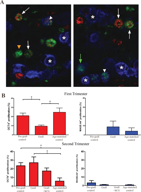
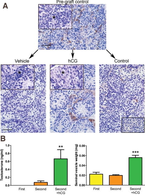
Human fetal testes (at 9 weeks, n = 4 and 14-18 weeks gestation, n = 6) were xenografted into male nude mice for 6 weeks, with or without hCG treatment of the host, and evaluated for normal cellular development and function using immunohistochemistry, triple immunofluorescence and testosterone assay. The differentiation and proliferation status of germ cells within xenografts was quantified and compared with age-matched controls.
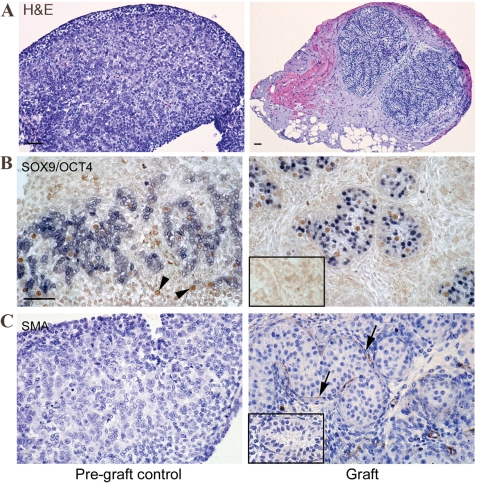
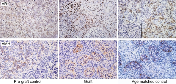
Xenografts showed >75% survival with normal morphology. In the first-trimester xenografts seminiferous cord formation was initiated and in first- and second-trimester grafts normal functional development of Sertoli, Leydig and peritubular myoid cells was demonstrated using cell-specific protein markers. Grafts produced testosterone when hosts were treated with hCG (P = 0.004 versus control). Proliferation of germ cells and differentiation from gonocytes (OCT4(+)) into pre-spermatogonia (VASA(+)) occurred in grafts and quantification showed this progressed comparably with age-matched ungrafted controls.
Human fetal testis tissue xenografts demonstrate normal structure, function and development after xenografting, including normal germ cell differentiation. This provides an in vivo system to study normal human fetal testis development and its susceptibility to disruption by exogenous factors (e.g. environmental chemicals). This should provide mechanistic insight into the fetal origins of DSDs and TDS disorders.
异常的胎儿睾丸发育可导致性别发育障碍(DSD),并易患睾丸发育不良综合征(TDS)疾病,如睾丸生殖细胞肿瘤。由于缺乏合适的模型和干预系统,人类胎儿睾丸发育的研究受到阻碍。我们假设人类胎儿睾丸异种移植物可以重现正常的发育。
将人类胎儿睾丸(9 周龄,n=4;14-18 周龄,n=6)异种移植到雄性裸鼠体内 6 周,并用或不用 hCG 处理宿主,并用免疫组织化学、三重免疫荧光和睾酮测定法评估正常的细胞发育和功能。定量比较异种移植物内生殖细胞的分化和增殖状态与年龄匹配的对照组。
异种移植物的存活率超过 75%,形态正常。在第一孕期的异种移植物中,开始形成精曲小管,在第一和第二孕期的移植物中,使用细胞特异性蛋白标志物证实了 Sertoli、Leydig 和小管周肌样细胞的正常功能发育。当宿主用 hCG 处理时,移植物产生睾酮(P=0.004 与对照组相比)。生殖细胞增殖并从性母细胞(OCT4(+))分化为精原细胞(VASA(+))发生在移植物中,定量分析显示其进展与未移植的年龄匹配对照组相当。
人类胎儿睾丸组织异种移植物在异种移植后表现出正常的结构、功能和发育,包括正常的生殖细胞分化。这为研究正常人类胎儿睾丸发育及其对外源性因素(如环境化学物质)易感性提供了体内系统。这应该为 DSD 和 TDS 疾病的胎儿起源提供机制上的见解。