MRC Centre for Reproductive Health, The Queen's Medical Research Institute, The University of Edinburgh, 47 Little France Crescent, Edinburgh, Scotland, UK.
School of Environmental and Life Sciences, Faculty of Science, University of Newcastle, Callaghan, NSW, Australia.
Hum Reprod. 2018 Nov 1;33(11):2107-2121. doi: 10.1093/humrep/dey289.
Does loss of DMRT1 in human fetal testis alter testicular development and result in testicular dysgenesis?
DMRT1 repression in human fetal testis alters the expression of key testicular and ovarian determining genes, and leads to focal testicular dysgenesis.
Testicular dysgenesis syndrome (TDS) is associated with common testicular disorders in young men, but its etiology is unknown. DMRT1 has been shown to play a role in the regulation of sex differentiation in the vertebrate gonad. Downregulation of DMRT1 in male mice results in trans-differentiation of Sertoli cells into granulosa (FOXL2+) cells resulting in an ovarian gonadal phenotype.
STUDY DESIGN, SIZE, DURATION: To determine the effect of DMRT1 repression on human fetal testes, we developed a novel system for genetic manipulation, which utilizes a Lentivral delivered miRNA during short-term in vitro culture (2 weeks). A long-term (4-6 weeks) ex vivo xenograft model was used to determine the subsequent effects of DMRT1 repression on testicular development and maintenance. We included first and second-trimester testis tissue (8-20 weeks gestation; n = 12) in the study.
PARTICIPANTS/MATERIALS, SETTING, METHODS: Human fetal testes were cultured in vitro and exposed to either of two DMRT1 miRNAs (miR536, miR641), or to scrambled control miRNA, for 24 h. This was followed by a further 14 days of culture (n = 3-4), or xenografting (n = 5) into immunocompromised mice for 4-6 weeks. Tissues were analyzed by histology, immunohistochemistry, immunofluorescence and quantitative RT-PCR. Endpoints included histological evaluation of seminiferous cord integrity, mRNA expression of testicular, ovarian and germ cell genes, and assessment of cell number and protein expression for proliferation, apoptosis and pluripotency factors. Statistical analysis was performed using a linear mixed effect model.
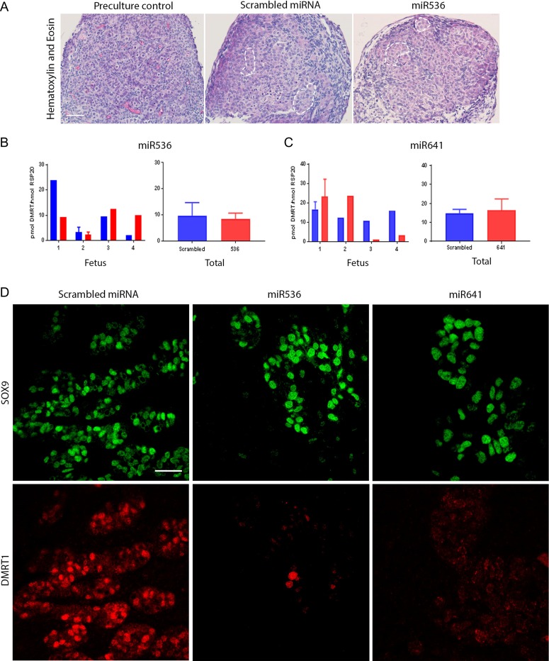
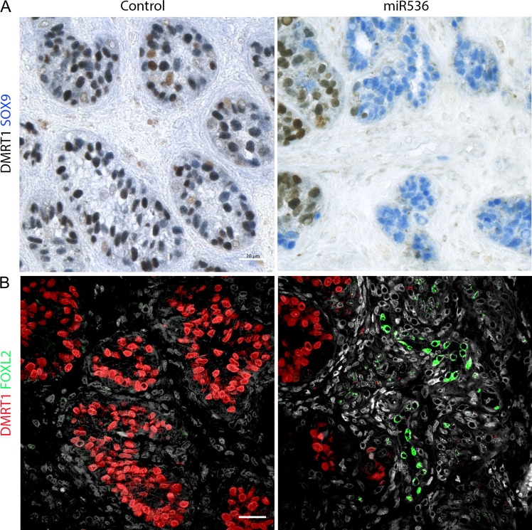
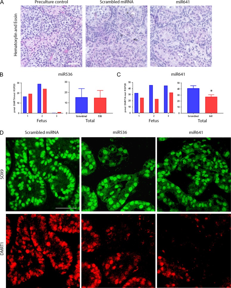
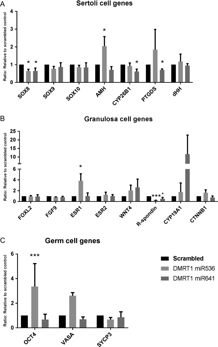
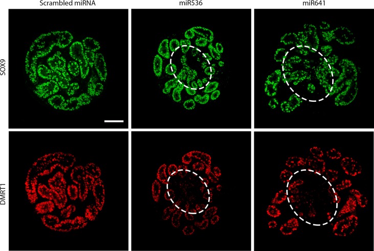
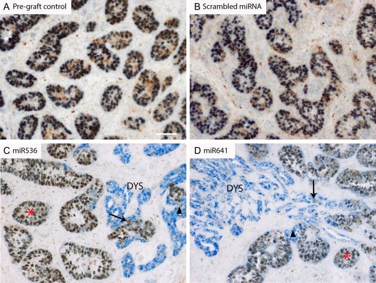
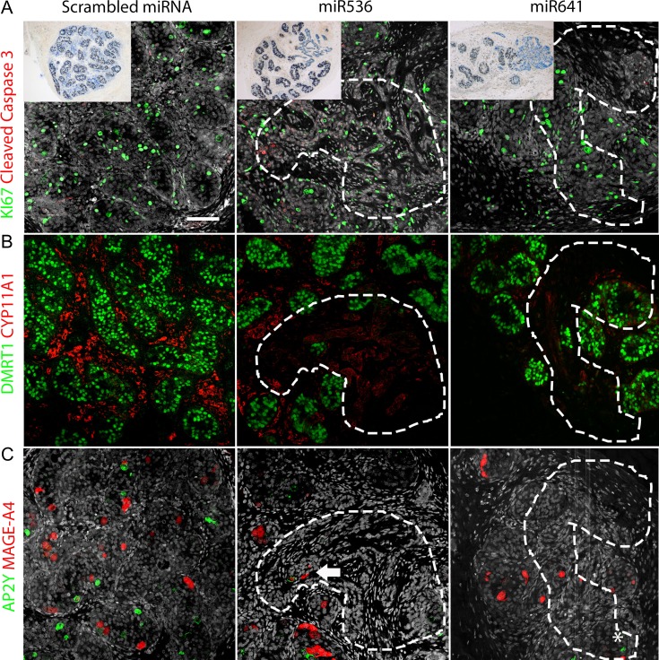
DMRT1 repression (miR536/miR641) resulted in a loss of DMRT1 protein expression in a sub-population of Sertoli cells of first trimester (8-11 weeks gestation) human fetal testis; however, this did not affect the completion of seminiferous cord formation or morphological appearance. In second-trimester testis (12-20 weeks gestation), DMRT1 repression (miR536/miR641) resulted in disruption of seminiferous cords with absence of DMRT1 protein expression in Sertoli (SOX9+) cells. No differences in proliferation (Ki67+) were observed and apoptotic cells (CC3+) were rare. Expression of the Sertoli cell associated gene, SOX8, was significantly reduced (miR536, 34% reduction, P = 0.031; miR641 36% reduction, P = 0.026), whilst SOX9 expression was unaffected. Changes in expression of AMH (miR536, 100% increase, P = 0.033), CYP26B1 (miR641, 38% reduction, P = 0.05) and PTGDS (miR642, 30% reduction, P = 0.0076) were also observed. Amongst granulosa cell associated genes, there was a significant downregulation in R-spondin 1 expression (miR536, 76% reduction, P < 0.0001; miR641, 49% reduction, P = 0.046); however, there were no changes in expression of the granulosa cell marker, FOXL2. Analysis of germ cell associated genes demonstrated a significant increase in the expression of the pluripotency gene OCT4 (miR536, 233%, P < 0.001). We used the xenograft system to investigate the longer-term effects of seminiferous cord disruption via DMRT1 repression. As was evident in vitro for second-trimester samples, DMRT1 repression resulted in focal testicular dysgenesis similar to that described in adults with TDS. These dysgenetic areas were devoid of germ cells, whilst expression of FOXL2 within the dysgenetic areas, indicated trans-differentiation from a male (Sertoli cell) to female (granulosa cell) phenotype.
LIMITATIONS, REASONS FOR CAUTION: Human fetal testis tissue is a limited resource; however, we were able to demonstrate significant effects of DMRT1 repression on the expression of germ and somatic cell genes, in addition to the induction of focal testicular dysgenesis, using these limited samples. In vitro culture may not reflect all aspects of human fetal testis development and function; however, the concurrent use of the xenograft model which represents a more physiological system supports the validity of the in vitro findings.
Our findings have important implications for understanding the role of DMRT1 in human testis development and in the origin of testicular dysgenesis. In addition, we provide validation of a novel system that can be used to determine the effects of repression of genes that have been implicated in gonadal development and associated human reproductive disorders.
STUDY FUNDING/COMPETING INTEREST(S): This project was funded by a Wellcome Trust Intermediate Clinical Fellowship (Grant No. 098522) awarded to RTM. LBS was supported by MRC Programme Grant MR/N002970/1. RAA was supported by MRC Programme Grant G1100357/1. RMS was supported by MRC Programme Grant G33253. This work was undertaken in the MRC Centre for Reproductive Health which is funded by the MRC Centre grant MR/N022556/1. The funding bodies had no input into the conduct of the research or the production of this manuscript. The authors have declared no conflicts of interest.
人类胎儿睾丸中 DMRT1 的缺失是否会改变睾丸发育并导致睾丸发育不良?
人类胎儿睾丸中 DMRT1 的抑制改变了关键的睾丸和卵巢决定基因的表达,并导致局灶性睾丸发育不良。
睾丸发育不良综合征(TDS)与年轻男性常见的睾丸疾病有关,但病因尚不清楚。DMRT1 已被证明在脊椎动物性腺的性别分化中发挥作用。在雄性小鼠中下调 DMRT1 会导致支持细胞向颗粒细胞(FOXL2+)的转分化,导致卵巢性腺表型。
研究设计、大小、持续时间:为了确定 DMRT1 抑制对人类胎儿睾丸的影响,我们开发了一种新的遗传操作系统,该系统利用短暂的体外培养(2 周)期间的 Lentivral 传递 miRNA。使用长期(4-6 周)异种移植模型来确定 DMRT1 抑制对睾丸发育和维持的后续影响。我们包括 8-20 周妊娠(n=12)的第一和第二孕期睾丸组织进行研究。
参与者/材料、设置、方法:将人类胎儿睾丸进行体外培养并暴露于两种 DMRT1 miRNA(miR536、miR641)中的一种或对照 miRNA 中 24 小时。接下来是进一步的 14 天培养(n=3-4),或 4-6 周的异种移植到免疫缺陷小鼠中。使用组织学、免疫组织化学、免疫荧光和定量 RT-PCR 分析组织。终点包括生精索完整性的组织学评估、睾丸、卵巢和生殖细胞基因的 mRNA 表达,以及增殖、凋亡和多能性因子的细胞数量和蛋白表达评估。使用线性混合效应模型进行统计分析。
DMRT1 抑制(miR536/miR641)导致第一孕期(8-11 周妊娠)人类胎儿睾丸中一小部分支持细胞中的 DMRT1 蛋白表达缺失;然而,这并没有影响生精索形成或形态的完成。在第二孕期睾丸(12-20 周妊娠)中,DMRT1 抑制(miR536/miR641)导致生精索中断,支持细胞(SOX9+)中缺失 DMRT1 蛋白表达。观察到增殖(Ki67+)没有差异,凋亡细胞(CC3+)很少。Sertoli 细胞相关基因 SOX8 的表达显著降低(miR536,减少 34%,P=0.031;miR641,减少 36%,P=0.026),而 SOX9 表达不受影响。AMH(miR536,增加 100%,P=0.033)、CYP26B1(miR641,减少 38%,P=0.05)和 PTGDS(miR642,减少 30%,P=0.0076)的表达也发生了变化。在颗粒细胞相关基因中,R-spondin 1 的表达显著下调(miR536,减少 76%,P<0.0001;miR641,减少 49%,P=0.046);然而,颗粒细胞标记物 FOXL2 的表达没有变化。生殖细胞相关基因的分析表明,多能性基因 OCT4 的表达显著增加(miR536,增加 233%,P<0.001)。我们使用异种移植系统来研究通过 DMRT1 抑制导致生精索中断的长期影响。正如第二孕期样本在体外观察到的那样,DMRT1 抑制导致局灶性睾丸发育不良类似于成人 TDS。这些发育不良区域缺乏生殖细胞,而在发育不良区域内 FOXL2 的表达表明从男性(支持细胞)到女性(颗粒细胞)表型的转分化。
局限性、谨慎的原因:人类胎儿睾丸组织是一种有限的资源;然而,我们能够使用这些有限的样本证明 DMRT1 抑制对生殖和体细胞基因表达的显著影响,以及诱导局灶性睾丸发育不良。体外培养可能无法反映人类胎儿睾丸发育和功能的所有方面;然而,同时使用代表更生理系统的异种移植模型支持了体外发现的有效性。
我们的研究结果对理解 DMRT1 在人类睾丸发育和睾丸发育不良起源中的作用具有重要意义。此外,我们提供了一种新系统的验证,该系统可用于确定已被认为与性腺发育和相关人类生殖障碍相关的基因抑制的影响。
研究资金/利益冲突:本项目由惠康信托中级临床奖学金(Grant No. 098522)资助,授予 RTM。LBS 得到 MRC 项目资助 MR/N002970/1 的支持。RAA 得到 MRC 项目资助 G1100357/1 的支持。RMS 得到 MRC 项目资助 G33253 的支持。这项工作是在 MRC 生殖健康中心进行的,该中心由 MRC 中心赠款 MR/N022556/1 资助。资助机构对研究的进行或本手稿的制作没有任何投入。作者没有声明利益冲突。