• 文献检索
  • 文档翻译
  • 深度研究
  • 学术资讯
  • Suppr Zotero 插件Zotero 插件
  • 邀请有礼
  • 套餐&价格
  • 历史记录
应用&插件
Suppr Zotero 插件Zotero 插件浏览器插件Mac 客户端Windows 客户端微信小程序
定价
高级版会员购买积分包购买API积分包
服务
文献检索文档翻译深度研究API 文档MCP 服务
关于我们
关于 Suppr公司介绍联系我们用户协议隐私条款
关注我们

Suppr 超能文献

核心技术专利:CN118964589B侵权必究
粤ICP备2023148730 号-1Suppr @ 2026

文献检索

告别复杂PubMed语法,用中文像聊天一样搜索,搜遍4000万医学文献。AI智能推荐,让科研检索更轻松。

立即免费搜索

文件翻译

保留排版,准确专业,支持PDF/Word/PPT等文件格式,支持 12+语言互译。

免费翻译文档

深度研究

AI帮你快速写综述,25分钟生成高质量综述,智能提取关键信息,辅助科研写作。

立即免费体验

儿童圆形肺炎及类似病变的影像学表现。

Imaging of round pneumonia and mimics in children.

机构信息

Department of Radiology, Miami Children's Hospital, Miami, FL 33155, USA.

出版信息

Pediatr Radiol. 2010 Dec;40(12):1931-40. doi: 10.1007/s00247-010-1767-7. Epub 2010 Aug 5.

DOI:10.1007/s00247-010-1767-7
PMID:20686763
原文链接:https://pmc.ncbi.nlm.nih.gov/articles/PMC7080009/
Abstract

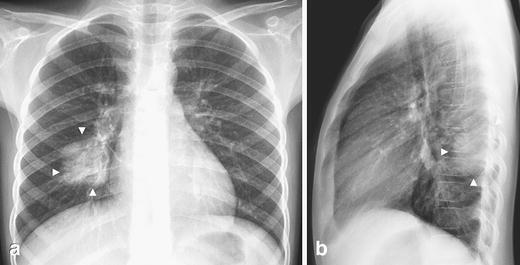
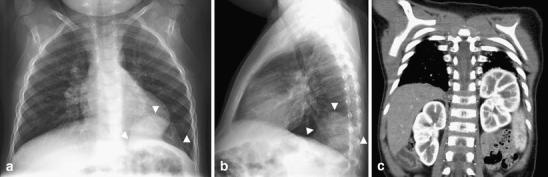
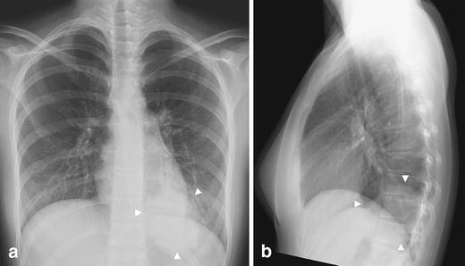
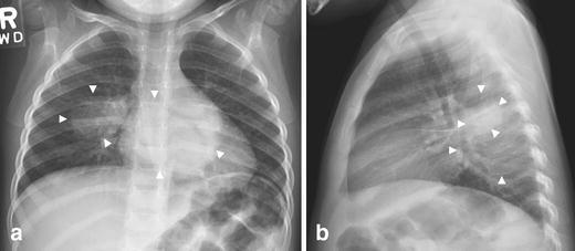
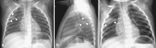
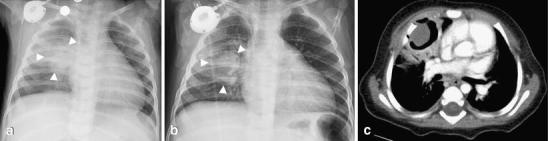
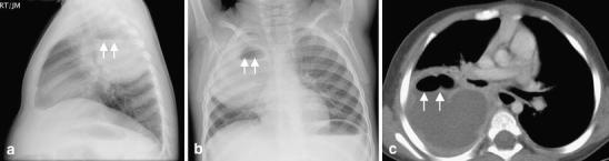
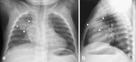
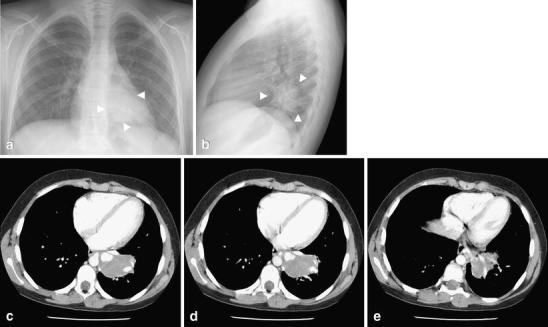
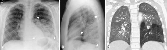
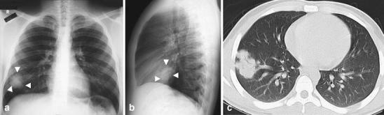
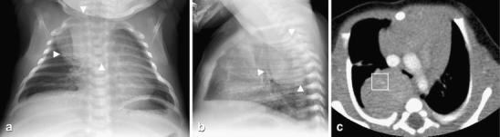
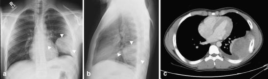
Various diseases in the pediatric age group can present as an intrathoracic rounded opacity on a chest radiograph. The purpose of this pictorial essay is to emphasize the imaging appearance of round pneumonia, an entity that occurs especially in the pediatric population. Additional pathologies with similar chest radiographic appearances are also presented. The diagnosis of round pneumonia should be made in children who have the typical clinical presentation along with chest radiographs demonstrating the characteristic findings.

摘要

在儿科年龄组中,各种疾病在胸部 X 光片上可表现为胸腔内圆形不透明影。本文旨在强调儿童中尤其常见的一种实体,即圆形肺炎的影像学表现。还介绍了具有类似胸部 X 线表现的其他病变。对于具有典型临床表现且胸部 X 光片显示特征性表现的儿童,应诊断为圆形肺炎。

https://cdn.ncbi.nlm.nih.gov/pmc/blobs/d85f/7080009/c71d9d240729/247_2010_1767_Fig13_HTML.jpg
https://cdn.ncbi.nlm.nih.gov/pmc/blobs/d85f/7080009/07c94aef18ed/247_2010_1767_Fig1_HTML.jpg
https://cdn.ncbi.nlm.nih.gov/pmc/blobs/d85f/7080009/5db27111e7d0/247_2010_1767_Fig2_HTML.jpg
https://cdn.ncbi.nlm.nih.gov/pmc/blobs/d85f/7080009/aff425dac4b7/247_2010_1767_Fig3_HTML.jpg
https://cdn.ncbi.nlm.nih.gov/pmc/blobs/d85f/7080009/3524bb214908/247_2010_1767_Fig4_HTML.jpg
https://cdn.ncbi.nlm.nih.gov/pmc/blobs/d85f/7080009/4d1cad94d867/247_2010_1767_Fig5_HTML.jpg
https://cdn.ncbi.nlm.nih.gov/pmc/blobs/d85f/7080009/18d1f57e2d77/247_2010_1767_Fig6_HTML.jpg
https://cdn.ncbi.nlm.nih.gov/pmc/blobs/d85f/7080009/03736c279ab8/247_2010_1767_Fig7_HTML.jpg
https://cdn.ncbi.nlm.nih.gov/pmc/blobs/d85f/7080009/c8767ffb2fc6/247_2010_1767_Fig8_HTML.jpg
https://cdn.ncbi.nlm.nih.gov/pmc/blobs/d85f/7080009/4388ff8e6bec/247_2010_1767_Fig9_HTML.jpg
https://cdn.ncbi.nlm.nih.gov/pmc/blobs/d85f/7080009/2b1386e40a00/247_2010_1767_Fig10_HTML.jpg
https://cdn.ncbi.nlm.nih.gov/pmc/blobs/d85f/7080009/9735f75bafec/247_2010_1767_Fig11_HTML.jpg
https://cdn.ncbi.nlm.nih.gov/pmc/blobs/d85f/7080009/f44d10c41b07/247_2010_1767_Fig12_HTML.jpg
https://cdn.ncbi.nlm.nih.gov/pmc/blobs/d85f/7080009/c71d9d240729/247_2010_1767_Fig13_HTML.jpg

相似文献

1
Imaging of round pneumonia and mimics in children.儿童圆形肺炎及类似病变的影像学表现。
Pediatr Radiol. 2010 Dec;40(12):1931-40. doi: 10.1007/s00247-010-1767-7. Epub 2010 Aug 5.
2
Computer-aided diagnosis for World Health Organization-defined chest radiograph primary-endpoint pneumonia in children.计算机辅助诊断在世界卫生组织定义的儿童胸片主要终点肺炎中的应用。
Pediatr Radiol. 2020 Apr;50(4):482-491. doi: 10.1007/s00247-019-04593-0. Epub 2020 Jan 13.
3
Pseudolesion of the chest. A conglomerate shadow on the lateral radiograph.
Chest. 1985 Apr;87(4):541-3. doi: 10.1378/chest.87.4.541.
4
Evaluation of pneumonia in children: comparison of MRI with fast imaging sequences at 1.5T with chest radiographs.儿童肺炎的评估:1.5T 快速成像序列磁共振成像与胸部 X 光片的比较
Acta Radiol. 2011 Oct 1;52(8):914-9. doi: 10.1258/ar.2011.100429. Epub 2011 Aug 4.
5
[Noninfectious differential diagnoses of pneumonia].[肺炎的非感染性鉴别诊断]
Radiologe. 2017 Jan;57(1):35-42. doi: 10.1007/s00117-016-0196-5.
6
Is the frontal radiograph alone sufficient to evaluate for pneumonia in children?仅胸部正位片就足以评估儿童是否患有肺炎吗?
Pediatr Radiol. 2004 May;34(5):379-83. doi: 10.1007/s00247-003-1128-x. Epub 2004 Jan 22.
7
A radiologic update on medical diseases of the newborn chest.
Pediatr Radiol. 1995;25(8):631-7. doi: 10.1007/BF02011835.
8
Randomised controlled trial of clinical outcome after chest radiograph in ambulatory acute lower-respiratory infection in children.儿童门诊急性下呼吸道感染胸部X光检查后临床结局的随机对照试验
Lancet. 1998 Feb 7;351(9100):404-8. doi: 10.1016/S0140-6736(97)07013-X.
9
Does the lateral chest radiograph help pediatric emergency physicians diagnose pneumonia? A randomized clinical trial.侧位胸部X光片对儿科急诊医生诊断肺炎有帮助吗?一项随机临床试验。
Acad Emerg Med. 2004 Jun;11(6):625-9.
10
Radiographic Imaging of Community-Acquired Pneumonia: A Case-Based Review.社区获得性肺炎的影像学表现:基于病例的综述。
Radiol Clin North Am. 2022 May;60(3):371-381. doi: 10.1016/j.rcl.2022.01.011.

引用本文的文献

1
ESR Essentials: imaging of common paediatric pulmonary diseases-practice recommendations by the European Society of Paediatric Radiology.红细胞沉降率要点:常见儿科肺部疾病的影像学检查——欧洲儿科放射学会的实践建议
Eur Radiol. 2025 Jan 29. doi: 10.1007/s00330-024-11268-4.
2
Solitary pulmonary nodule in an early adolescent.青少年早期孤立性肺结节。
BMJ Case Rep. 2023 Sep 11;16(9):e256081. doi: 10.1136/bcr-2023-256081.
3
Unilateral double round pneumonia in a child: A case report and literature review.儿童单侧双圆形肺炎:一例报告及文献综述

本文引用的文献

1
Right-sided Bochdalek Hernia Presenting as a Solitary Pulmonary Nodule.表现为孤立性肺结节的右侧博赫dalek疝
Radiol Case Rep. 2015 Dec 7;3(2):114. doi: 10.2484/rcr.v3i2.114. eCollection 2008.
2
Bronchopulmonary foregut malformations presenting as mass lesions in children: spectrum of imaging findings.支气管肺前肠畸形表现为儿童肿块病变:影像学表现谱。
Diagn Interv Radiol. 2010 Jun;16(2):153-61. doi: 10.4261/1305-3825.DIR.2063-08.2. Epub 2009 Oct 9.
3
CT imaging of mass-like nonvascular pulmonary lesions in children.儿童肺部类肿块非血管性病变的CT成像
Radiol Case Rep. 2021 Aug 26;16(11):3266-3269. doi: 10.1016/j.radcr.2021.07.066. eCollection 2021 Nov.
4
The Echo of Pulmonary Tuberculosis: Mechanisms of Clinical Symptoms and Other Disease-Induced Systemic Complications.肺结核的回响:临床症状的发生机制及其他疾病引发的全身性并发症。
Clin Microbiol Rev. 2020 Jul 1;33(4). doi: 10.1128/CMR.00036-20. Print 2020 Sep 16.
5
Round pneumonia due to in a child.一名儿童因[病因未提及]导致的圆形肺炎。
Radiol Case Rep. 2019 Jan 18;14(4):436-438. doi: 10.1016/j.radcr.2019.01.010. eCollection 2019 Apr.
6
[Specific characteristics of chest X‑ray in childhood : Basics for radiologists].[儿童胸部X线的特殊特征:放射科医生的基础]
Radiologe. 2018 Apr;58(4):359-376. doi: 10.1007/s00117-018-0374-8.
7
Assessment of chest X-ray images in newborns with respiratory disorders.患有呼吸系统疾病的新生儿胸部X光图像评估。
Kardiochir Torakochirurgia Pol. 2015 Mar;12(1):83-6. doi: 10.5114/kitp.2015.50578. Epub 2015 Mar 31.
8
Pediatric round pneumonia.小儿圆形肺炎
Pediatr Neonatol. 2014 Dec;55(6):491-4. doi: 10.1016/j.pedneo.2013.01.014. Epub 2013 Mar 13.
9
Single pulmonary round mass in a 12-year-old boy.一名12岁男孩的单个肺圆形肿块。
BMJ Case Rep. 2012 Jul 3;2012:bcr2012006356. doi: 10.1136/bcr-2012-006356.
Pediatr Radiol. 2007 Dec;37(12):1253-63. doi: 10.1007/s00247-007-0637-4. Epub 2007 Oct 31.
4
Round pneumonia: imaging findings in a large series of children.圆形肺炎:大量儿童的影像学表现
Pediatr Radiol. 2007 Dec;37(12):1235-40. doi: 10.1007/s00247-007-0654-3. Epub 2007 Oct 19.
5
Fungal infections in primary immunodeficiencies.原发性免疫缺陷中的真菌感染
Eur J Pediatr. 2007 Nov;166(11):1099-117. doi: 10.1007/s00431-007-0527-7. Epub 2007 Jun 6.
6
Imaging findings in acute invasive pulmonary aspergillosis: clinical significance of the halo sign.急性侵袭性肺曲霉病的影像学表现:晕征的临床意义。
Clin Infect Dis. 2007 Feb 1;44(3):373-9. doi: 10.1086/509917. Epub 2006 Dec 29.
7
Congenital bronchopulmonary foregut malformations: concepts and controversies.先天性支气管肺前肠畸形:概念与争议
Pediatr Radiol. 2006 Aug;36(8):773-91. doi: 10.1007/s00247-006-0115-4. Epub 2006 Mar 22.
8
One-month-old infant with multilobar round pneumonias.
Pediatr Infect Dis J. 2006 Jan;25(1):95, 97. doi: 10.1097/01.inf.0000195717.40250.8b.
9
Neuroblastoma in childhood: review and radiological findings.儿童神经母细胞瘤:综述与影像学表现
Cancer Imaging. 2005 Sep 30;5(1):116-27. doi: 10.1102/1470-7330.2005.0104.
10
Imaging in immunocompetent children who have pneumonia.免疫功能正常的肺炎患儿的影像学检查
Radiol Clin North Am. 2005 Mar;43(2):253-65. doi: 10.1016/j.rcl.2004.11.001.