Biomedical NMR, Department of Biomedical Engineering, Eindhoven University of Technology, Eindhoven, The Netherlands.
Bioconjug Chem. 2010 Oct 20;21(10):1794-803. doi: 10.1021/bc100091q.
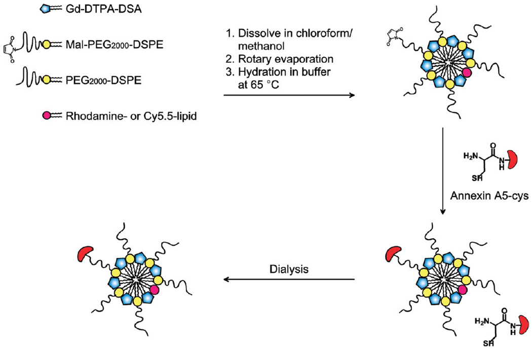
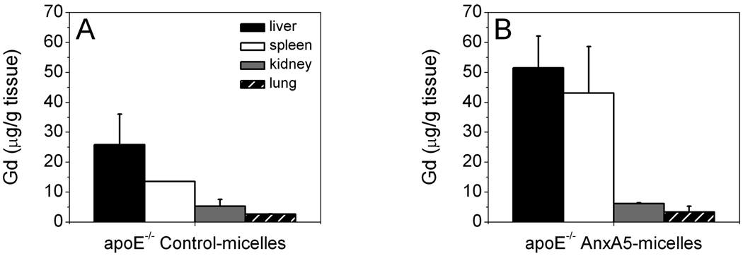
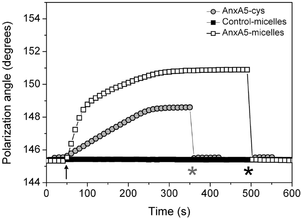
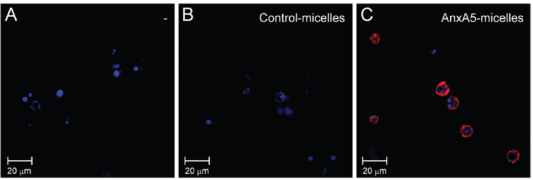
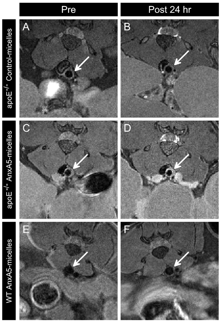
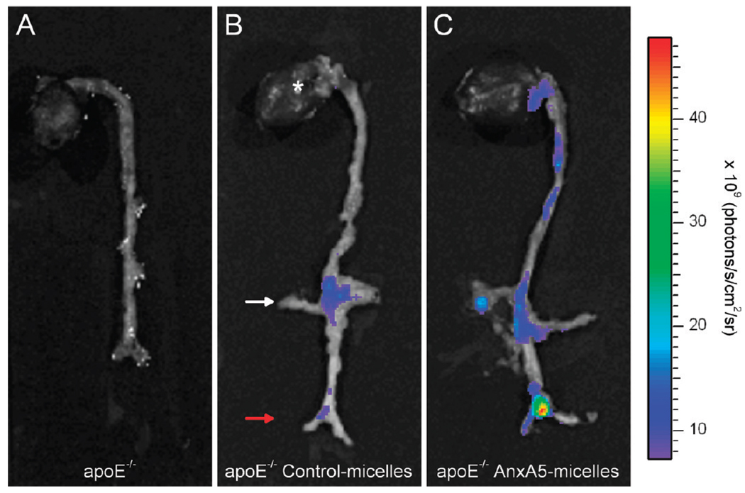
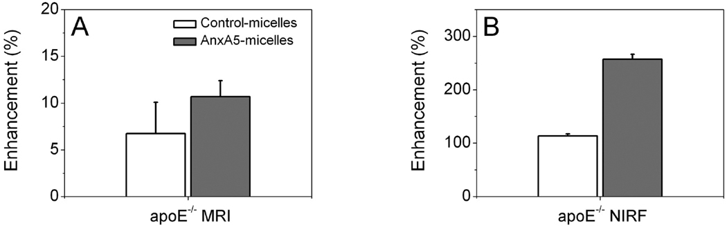
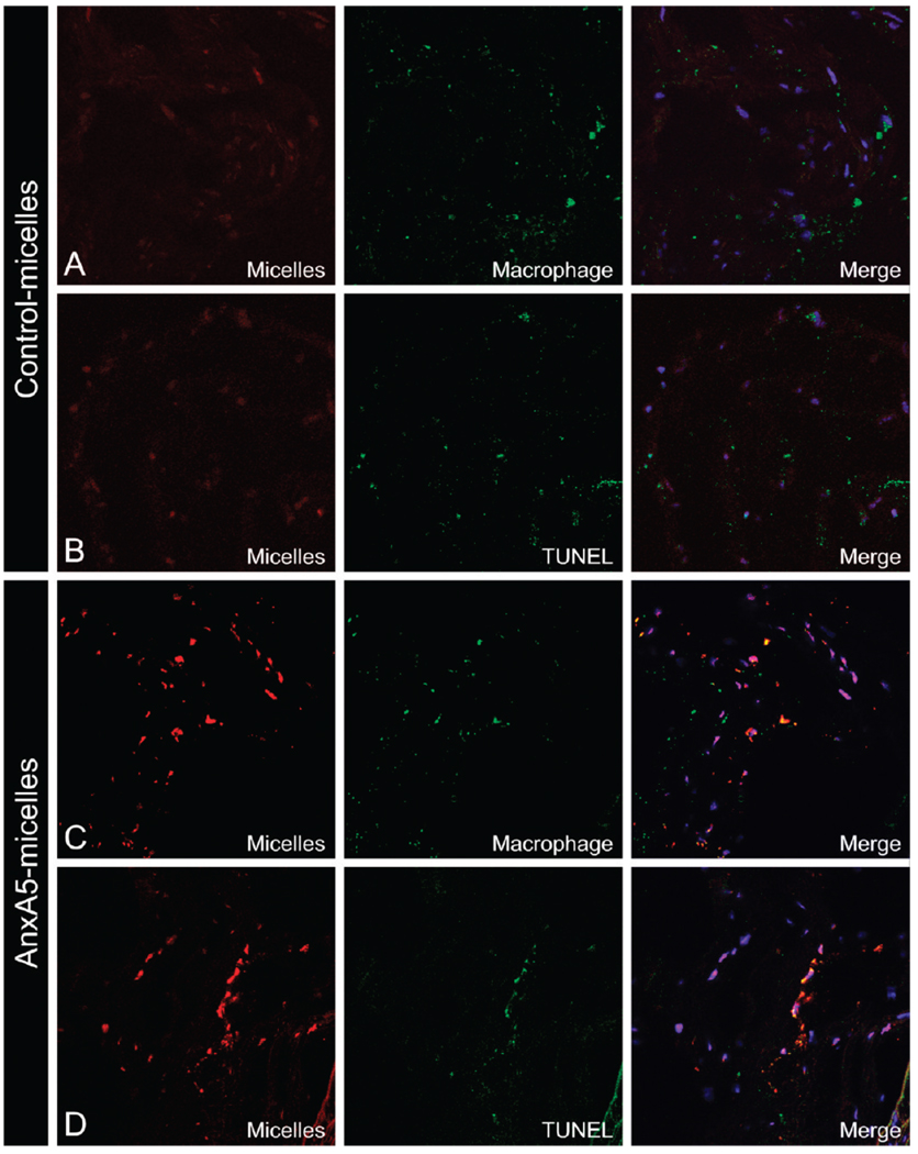
Apoptosis and macrophage burden are believed to correlate with atherosclerotic plaque vulnerability and are therefore considered important diagnostic and therapeutic targets for atherosclerosis. These cell types are characterized by the exposure of phosphatidylserine (PS) at their surface. In the present study, we developed and applied a small micellar fluorescent annexin A5-functionalized nanoparticle for noninvasive magnetic resonance imaging (MRI) of PS exposing cells in atherosclerotic lesions. Annexin A5-mediated target-specificity was confirmed with ellipsometry and in vitro binding to apoptotic Jurkat cells. In vivo T(1)-weighted MRI of the abdominal aorta in atherosclerotic ApoE(-/-) mice revealed enhanced uptake of the annexin A5-micelles as compared to control-micelles, which was corroborated with ex vivo near-infrared fluorescence images of excised whole aortas. Confocal laser scanning microscopy (CLSM) demonstrated that the targeted agent was associated with macrophages and apoptotic cells, whereas the nonspecific control agent showed no clear uptake by such cells. In conclusion, the annexin A5-conjugated bimodal micelles displayed potential for noninvasive assessment of cell types that are considered to significantly contribute to plaque instability and therefore may be of great value in the assessment of atherosclerotic lesion phenotype.
细胞凋亡和巨噬细胞负担被认为与动脉粥样硬化斑块的脆弱性相关,因此被认为是动脉粥样硬化的重要诊断和治疗靶点。这些细胞类型的特征是其表面暴露磷酸丝氨酸(PS)。在本研究中,我们开发并应用了一种小胶束荧光型 annexin A5 功能化纳米颗粒,用于动脉粥样硬化病变中 PS 暴露细胞的非侵入性磁共振成像(MRI)。通过椭圆术和体外与凋亡 Jurkat 细胞的结合,证实了 annexin A5 介导的靶向特异性。与对照胶束相比,在动脉粥样硬化 ApoE(-/-)小鼠的腹主动脉进行的体内 T1 加权 MRI 显示 annexin A5 胶束的摄取增强,这与切除的整个主动脉的近红外荧光图像的体外结果相符。共聚焦激光扫描显微镜(CLSM)显示,靶向剂与巨噬细胞和凋亡细胞相关,而非特异性对照剂则未显示出此类细胞的明显摄取。总之,这种 annexin A5 偶联的双模态胶束具有非侵入性评估被认为对斑块不稳定有重大贡献的细胞类型的潜力,因此在评估动脉粥样硬化病变表型方面可能具有重要价值。