Clark J M
Department of Orthopaedics, University of Washington, School of Medicine, Seattle 98195.
J Anat. 1990 Aug;171:105-15.
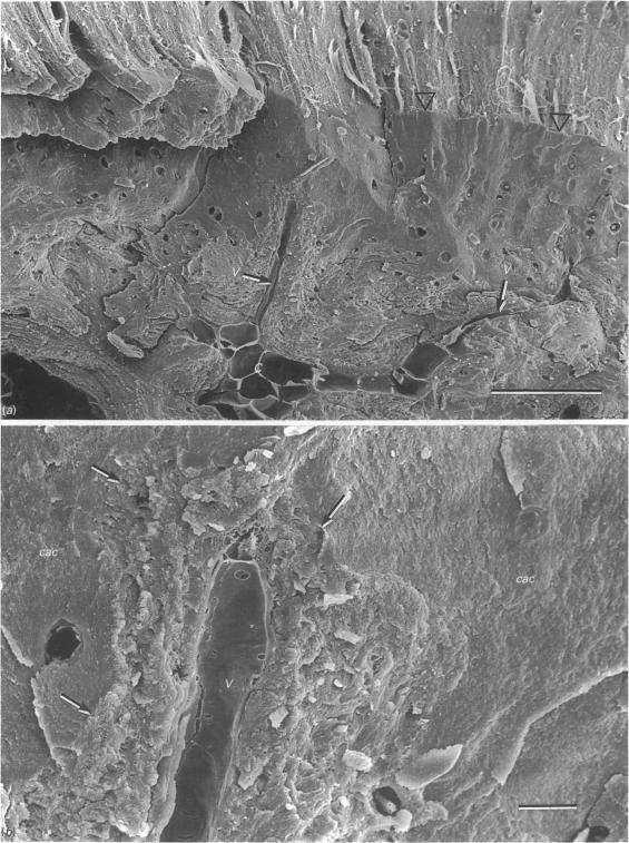
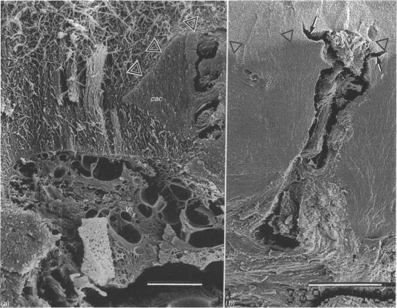
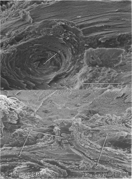
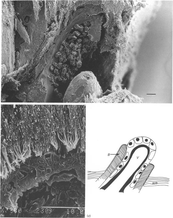
Human, rabbit and canine articular cartilage was prepared for SEM by fixation in isosmolal glutaraldehyde and freeze-fracture following dehydration. These techniques produced clear images of the bone, marrow and vessels in the subchondral region. Generally, cavities larger than 40 microns contained elements typical of marrow. Capillaries ran through the bone in cylindrical channels 20-40 microns wide. These channels were surrounded by concentric lamellae of bone and were in all respects Haversian canals within osteons. A minority of these channels opened into the calcified articular cartilage and there were preceded by cells which appeared to be cutting into the cartilage. Most vascular channels, however, were separated from the cartilage by a layer of bone. We conclude that the vessels within subchondral bone are present primarily to supply the bone through a network of mature osteons.
通过在等渗戊二醛中固定并在脱水后进行冷冻断裂,制备了用于扫描电子显微镜(SEM)观察的人、兔和犬的关节软骨。这些技术产生了软骨下区域骨骼、骨髓和血管的清晰图像。一般来说,大于40微米的腔隙包含典型的骨髓成分。毛细血管在宽20 - 40微米的圆柱形通道中穿过骨骼。这些通道被同心的骨板包围,在各方面都是骨单位内的哈弗斯管。这些通道中的少数通向钙化的关节软骨,并且在其之前有似乎切入软骨的细胞。然而,大多数血管通道被一层骨与软骨分隔开。我们得出结论,软骨下骨内的血管主要是通过成熟骨单位网络为骨骼提供营养。