Institute for Biology - Neurobiology, Free University Berlin Berlin, Germany.
Front Syst Neurosci. 2010 Jul 13;4. doi: 10.3389/fnsys.2010.00030. eCollection 2010.
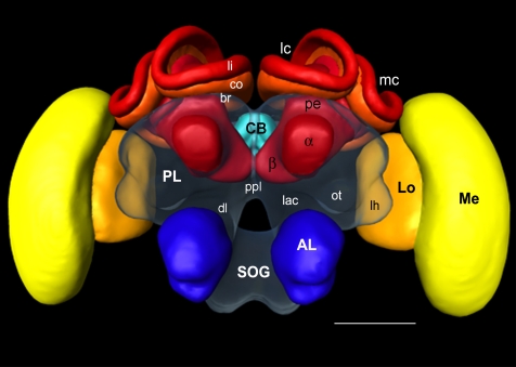
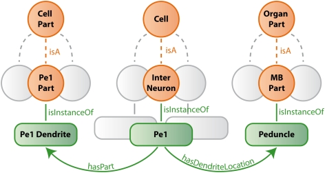
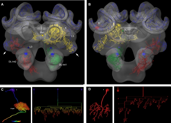
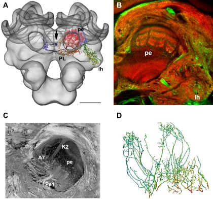
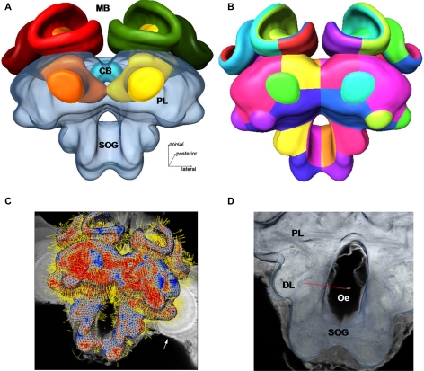
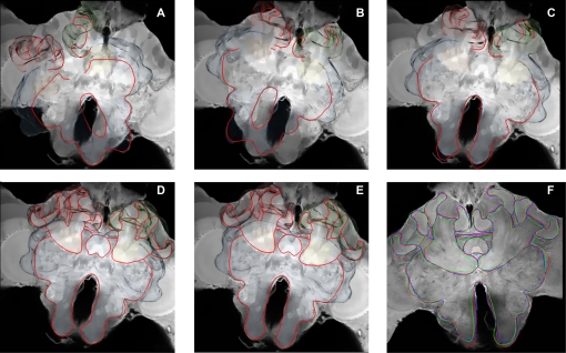
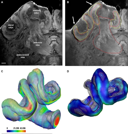
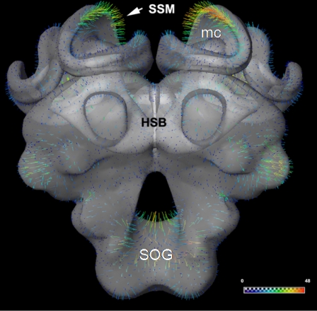
The honeybee standard brain (HSB) serves as an interactive tool for relating morphologies of bee brain neurons and provides a reference system for functional and bibliographical properties (http://www.neurobiologie.fu-berlin.de/beebrain/). The ultimate goal is to document not only the morphological network properties of neurons collected from separate brains, but also to establish a graphical user interface for a neuron-related data base. Here, we review the current methods and protocols used to incorporate neuronal reconstructions into the HSB. Our registration protocol consists of two separate steps applied to imaging data from two-channel confocal microscopy scans: (1) The reconstruction of the neuron, facilitated by an automatic extraction of the neuron's skeleton based on threshold segmentation, and (2) the semi-automatic 3D segmentation of the neuropils and their registration with the HSB. The integration of neurons in the HSB is performed by applying the transformation computed in step (2) to the reconstructed neurons of step (1). The most critical issue of this protocol in terms of user interaction time - the segmentation process - is drastically improved by the use of a model-based segmentation process. Furthermore, the underlying statistical shape models (SSM) allow the visualization and analysis of characteristic variations in large sets of bee brain data. The anatomy of neural networks composed of multiple neurons that are registered into the HSB are visualized by depicting the 3D reconstructions together with semantic information with the objective to integrate data from multiple sources (electrophysiology, imaging, immunocytochemistry, molecular biology). Ultimately, this will allow the user to specify cell types and retrieve their morphologies along with physiological characterizations.
蜜蜂标准脑(HSB)可用作相关蜜蜂脑神经元形态的交互工具,并提供功能和文献属性的参考系统(http://www.neurobiologie.fu-berlin.de/beebrain/)。最终目标不仅是记录从单独的大脑中收集的神经元的形态网络属性,还要为神经元相关数据库建立图形用户界面。在这里,我们回顾了将神经元重建纳入 HSB 中当前使用的方法和协议。我们的注册协议由两步组成,应用于双通道共聚焦显微镜扫描的成像数据:(1) 神经元的重建,基于阈值分割的自动提取神经元骨架来辅助完成,以及 (2) 神经节的半自动 3D 分割及其与 HSB 的配准。通过将步骤 (2) 中计算的变换应用于步骤 (1) 中的重建神经元来完成 HSB 中神经元的整合。就用户交互时间而言,此协议中最关键的问题——分割过程——通过使用基于模型的分割过程得到了极大的改善。此外,基础统计形状模型(SSM)允许对大量蜜蜂脑数据中的特征变化进行可视化和分析。通过绘制 3D 重建图以及具有语义信息的图来可视化由注册到 HSB 的多个神经元组成的神经网络的解剖结构,目的是整合来自多个来源的数据(电生理学、成像、免疫细胞化学、分子生物学)。最终,这将允许用户指定细胞类型并检索它们的形态以及生理特征。