• 文献检索
  • 文档翻译
  • 深度研究
  • 学术资讯
  • Suppr Zotero 插件Zotero 插件
  • 邀请有礼
  • 套餐&价格
  • 历史记录
应用&插件
Suppr Zotero 插件Zotero 插件浏览器插件Mac 客户端Windows 客户端微信小程序
定价
高级版会员购买积分包购买API积分包
服务
文献检索文档翻译深度研究API 文档MCP 服务
关于我们
关于 Suppr公司介绍联系我们用户协议隐私条款
关注我们

Suppr 超能文献

核心技术专利:CN118964589B侵权必究
粤ICP备2023148730 号-1Suppr @ 2026

文献检索

告别复杂PubMed语法,用中文像聊天一样搜索,搜遍4000万医学文献。AI智能推荐,让科研检索更轻松。

立即免费搜索

文件翻译

保留排版,准确专业,支持PDF/Word/PPT等文件格式,支持 12+语言互译。

免费翻译文档

深度研究

AI帮你快速写综述,25分钟生成高质量综述,智能提取关键信息,辅助科研写作。

立即免费体验

使用四维超声时空图像相关技术(STIC)的11个胎儿超声心动图平面:一种胎儿心脏容积分析的合理方法。

Eleven fetal echocardiographic planes using 4-dimensional ultrasound with spatio-temporal image correlation (STIC): a logical approach to fetal heart volume analysis.

作者信息

Jantarasaengaram Surasak, Vairojanavong Kittipong

机构信息

Maternal-Fetal Medicine Unit and Ultrasound Unit, Rajavithi Hospital, College of Medicine, Rangsit University, Bangkok, Thailand.

出版信息

Cardiovasc Ultrasound. 2010 Sep 15;8:41. doi: 10.1186/1476-7120-8-41.

DOI:10.1186/1476-7120-8-41
PMID:20843340
原文链接:https://pmc.ncbi.nlm.nih.gov/articles/PMC2949622/
Abstract

BACKGROUND

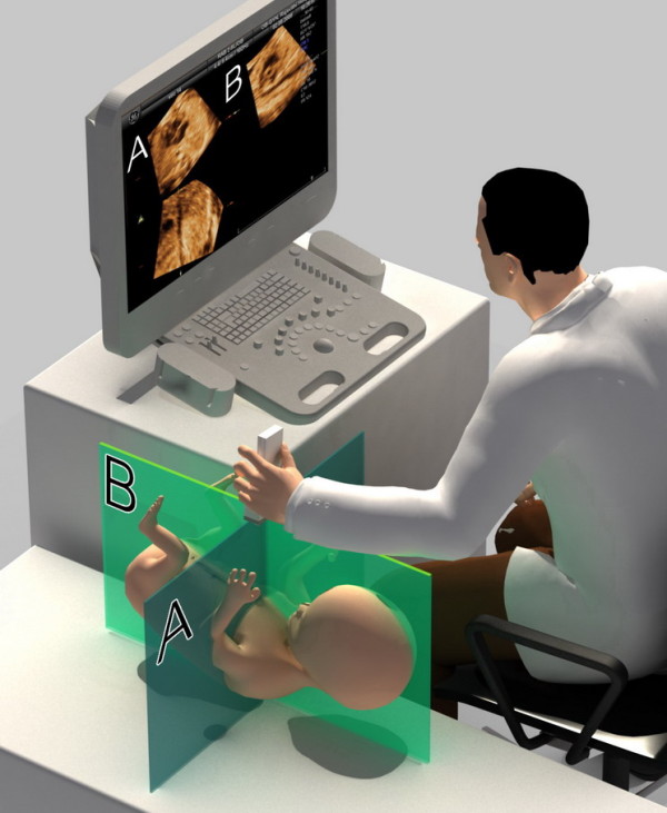
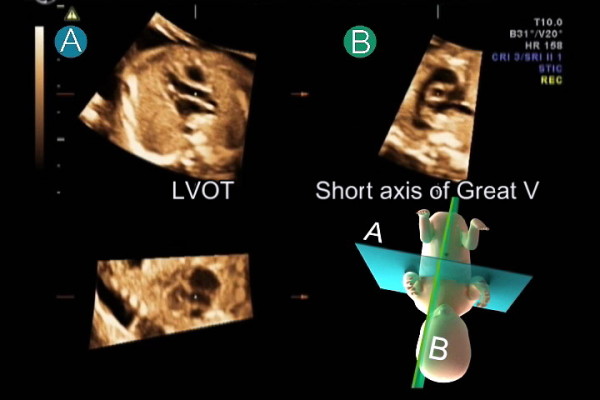
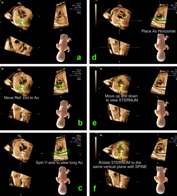
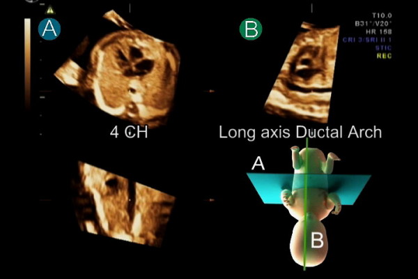
Theoretically, a cross-sectional image of any cardiac planes can be obtained from a STIC fetal heart volume dataset. We described a method to display 11 fetal echocardiographic planes from STIC volumes.

METHODS

Fetal heart volume datasets were acquired by transverse acquisition from 200 normal fetuses at 15 to 40 weeks of gestation. Analysis of the volume datasets using the described technique to display 11 echocardiographic planes in the multiplanar display mode were performed offline.

RESULTS

Volume datasets from 18 fetuses were excluded due to poor image resolution. The mean visualization rates for all echocardiographic planes at 15-17, 18-22, 23-27, 28-32 and 33-40 weeks of gestation fetuses were 85.6% (range 45.2-96.8%, N = 31), 92.9% (range 64.0-100%, N = 64), 93.4% (range 51.4-100%, N = 37), 88.7%(range 54.5-100%, N = 33) and 81.8% (range 23.5-100%, N = 17) respectively.

CONCLUSIONS

Overall, the applied technique can favorably display the pertinent echocardiographic planes. Description of the presented method provides a logical approach to explore the fetal heart volumes.

摘要

背景

从理论上讲,任何心脏平面的横截面图像都可以从STIC胎儿心脏容积数据集中获取。我们描述了一种从STIC容积中显示11个胎儿超声心动图平面的方法。

方法

对200例孕15至40周的正常胎儿进行横向采集,获取胎儿心脏容积数据集。使用所描述的技术对容积数据集进行分析,以在多平面显示模式下显示11个超声心动图平面,并进行离线分析。

结果

由于图像分辨率差,排除了18例胎儿的容积数据集。孕15 - 17周、18 - 22周、23 - 27周、28 - 32周和33 - 40周胎儿所有超声心动图平面的平均显示率分别为85.6%(范围45.2 - 96.8%,N = 31)、92.9%(范围64.0 - 100%,N = 64)、93.4%(范围51.4 - 100%,N = 37)、88.7%(范围54.5 - 100%,N = 33)和81.8%(范围23.5 - 100%,N = 17)。

结论

总体而言,所应用的技术能够很好地显示相关的超声心动图平面。所提出方法的描述为探索胎儿心脏容积提供了一种合理的方法。

https://cdn.ncbi.nlm.nih.gov/pmc/blobs/b71e/2949622/04e2474b1f58/1476-7120-8-41-4.jpg
https://cdn.ncbi.nlm.nih.gov/pmc/blobs/b71e/2949622/5f4923fb8a92/1476-7120-8-41-1.jpg
https://cdn.ncbi.nlm.nih.gov/pmc/blobs/b71e/2949622/16d4f7b686c4/1476-7120-8-41-2.jpg
https://cdn.ncbi.nlm.nih.gov/pmc/blobs/b71e/2949622/0d49665c1c08/1476-7120-8-41-3.jpg
https://cdn.ncbi.nlm.nih.gov/pmc/blobs/b71e/2949622/04e2474b1f58/1476-7120-8-41-4.jpg

相似文献

1
Eleven fetal echocardiographic planes using 4-dimensional ultrasound with spatio-temporal image correlation (STIC): a logical approach to fetal heart volume analysis.使用四维超声时空图像相关技术(STIC)的11个胎儿超声心动图平面:一种胎儿心脏容积分析的合理方法。
Cardiovasc Ultrasound. 2010 Sep 15;8:41. doi: 10.1186/1476-7120-8-41.
2
Three-dimensional (3D) and 4D color Doppler fetal echocardiography using spatio-temporal image correlation (STIC).使用时空图像相关技术(STIC)的三维(3D)和四维(4D)彩色多普勒胎儿超声心动图。
Ultrasound Obstet Gynecol. 2004 Jun;23(6):535-45. doi: 10.1002/uog.1075.
3
Four-dimensional ultrasonography of the fetal heart using a novel Tomographic Ultrasound Imaging display.使用新型断层超声成像显示技术对胎儿心脏进行四维超声检查。
J Perinat Med. 2006;34(1):39-55. doi: 10.1515/JPM.2006.006.
4
Use of spatiotemporal image correlation at 11-14 weeks' gestation.使用时空关联成像技术在 11-14 孕周检查。
Ultrasound Obstet Gynecol. 2013 Dec;42(6):669-78. doi: 10.1002/uog.12548.
5
Four-chamber view and 'swing technique' (FAST) echo: a novel and simple algorithm to visualize standard fetal echocardiographic planes.四腔心切面和“摆动技术”(FAST)超声:一种新颖而简单的方法,用于可视化标准胎儿超声心动图平面。
Ultrasound Obstet Gynecol. 2011 Apr;37(4):423-31. doi: 10.1002/uog.8840. Epub 2011 Mar 2.
6
Fetal cardiac function assessed with four-dimensional ultrasound imaging using spatiotemporal image correlation.应用时空相关成像技术的四维超声对胎儿心功能的评估。
Ultrasound Obstet Gynecol. 2009 Mar;33(3):272-81. doi: 10.1002/uog.6287.
7
Four-dimensional ultrasonography of the fetal heart with spatiotemporal image correlation.胎儿心脏的四维超声检查与时空图像相关技术
Am J Obstet Gynecol. 2003 Dec;189(6):1792-802. doi: 10.1016/s0002-9378(03)00913-x.
8
Electronic versus conventional spatiotemporal image correlation (STIC) fetal echocardiography: a direct comparison.电子与传统时空关联(STIC)胎儿超声心动图:直接比较。
J Matern Fetal Neonatal Med. 2022 Apr;35(7):1310-1317. doi: 10.1080/14767058.2020.1752656. Epub 2020 Apr 15.
9
Fetal cardiac ventricle volumetry in the second half of gestation assessed by 4D ultrasound using STIC combined with inversion mode.孕晚期胎儿心室容积测量:采用 4D 超声 STIC 联合反转模式进行评估
Ultrasound Obstet Gynecol. 2007 Aug;30(2):142-51. doi: 10.1002/uog.4036.
10
Reproducibility of echocardiographic measurements of human fetal left ventricular volumes and ejection fractions using four-dimensional ultrasound with the spatio-temporal image correlation modality.应用时空关联模式的四维超声技术测量人胎儿左心室容量和射血分数的可重复性。
Eur J Obstet Gynecol Reprod Biol. 2012 Jan;160(1):22-9. doi: 10.1016/j.ejogrb.2011.09.036. Epub 2011 Oct 21.

引用本文的文献

1
Fetal intelligent navigation echocardiography (FINE) has superior performance compared to manual navigation of the fetal heart by non-expert sonologists.胎儿智能导航超声心动图(FINE)在非专家超声医师手动导航胎儿心脏方面表现更优。
J Perinat Med. 2022 Dec 7;51(4):477-491. doi: 10.1515/jpm-2022-0387. Print 2023 May 25.
2
Fetal Intelligent Navigation Echocardiography (FINE) Detects 98% of Congenital Heart Disease.胎儿智能导航超声心动图(FINE)可检测出98%的先天性心脏病。
J Ultrasound Med. 2018 Nov;37(11):2577-2593. doi: 10.1002/jum.14616. Epub 2018 Mar 30.
3
A Prospective Study of the Use of Fetal Intelligent Navigation Echocardiography (FINE) to Obtain Standard Fetal Echocardiography Views.

本文引用的文献

1
Cardiac screening by STIC: can sonologists performing the 20-week anomaly scan pick up outflow tract abnormalities by scrolling the A-plane of STIC volumes?通过时空关联成像(STIC)进行心脏筛查:进行20周异常扫描的超声科医生能否通过滚动STIC容积的A平面来发现流出道异常?
Ultrasound Obstet Gynecol. 2008 Dec;32(7):865-70. doi: 10.1002/uog.6261.
2
ISUOG consensus statement: what constitutes a fetal echocardiogram?国际妇产科超声学会共识声明:什么构成胎儿超声心动图?
Ultrasound Obstet Gynecol. 2008 Aug;32(2):239-42. doi: 10.1002/uog.6115.
3
A systematic analysis of the feasibility of four-dimensional ultrasound imaging using spatiotemporal image correlation in routine fetal echocardiography.
一项关于使用胎儿智能导航超声心动图(FINE)获取标准胎儿超声心动图视图的前瞻性研究。
Fetal Diagn Ther. 2017;41(2):89-99. doi: 10.1159/000446982. Epub 2016 Jun 17.
4
How to Acquire Cardiac Volumes for Sonographic Examination of the Fetal Heart: Part 1.如何获取胎儿心脏超声检查的心脏容积:第1部分。
J Ultrasound Med. 2016 May;35(5):1021-42. doi: 10.7863/ultra.16.01081. Epub 2016 Apr 18.
5
How to Acquire Cardiac Volumes for Sonographic Examination of the Fetal Heart: Part 2.如何获取用于胎儿心脏超声检查的心脏容积:第2部分。
J Ultrasound Med. 2016 May;35(5):1043-66. doi: 10.7863/ultra.16.01082. Epub 2016 Apr 18.
6
Intelligent navigation to improve obstetrical sonography.智能导航改善产科超声检查。
Ultrasound Obstet Gynecol. 2016 Apr;47(4):403-9. doi: 10.1002/uog.12562. Epub 2015 Nov 3.
7
Prospective evaluation of the fetal heart using Fetal Intelligent Navigation Echocardiography (FINE).使用胎儿智能导航超声心动图(FINE)对胎儿心脏进行前瞻性评估。
Ultrasound Obstet Gynecol. 2016 Apr;47(4):450-9. doi: 10.1002/uog.15676. Epub 2016 Mar 10.
8
Fetal Intelligent Navigation Echocardiography (FINE): a novel method for rapid, simple, and automatic examination of the fetal heart.胎儿智能导航超声心动图(FINE):一种快速、简便、自动检查胎儿心脏的新方法。
Ultrasound Obstet Gynecol. 2013 Sep;42(3):268-84. doi: 10.1002/uog.12563.
在常规胎儿超声心动图中使用时空图像相关技术进行四维超声成像可行性的系统分析。
Ultrasound Obstet Gynecol. 2008 Jun;31(6):625-32. doi: 10.1002/uog.5351.
4
Automated retrieval of standard diagnostic fetal cardiac ultrasound planes in the second trimester of pregnancy: a prospective evaluation of software.妊娠中期标准诊断性胎儿心脏超声平面的自动检索:软件的前瞻性评估
Ultrasound Obstet Gynecol. 2008 Jan;31(1):30-6. doi: 10.1002/uog.5228.
5
The role of the sagittal view of the ductal arch in identification of fetuses with conotruncal anomalies using 4-dimensional ultrasonography.导管弓矢状面在使用四维超声识别圆锥干畸形胎儿中的作用。
J Ultrasound Med. 2007 Sep;26(9):1181-8; quiz 1189-90. doi: 10.7863/jum.2007.26.9.1181.
6
Standardization of on-screen fetal heart orientation prior to storage of spatio-temporal image correlation (STIC) volume datasets.在存储时空图像相关(STIC)容积数据集之前对屏幕上胎儿心脏方向进行标准化。
Ultrasound Obstet Gynecol. 2007 Jun;29(6):605-11. doi: 10.1002/uog.4049.
7
Automated sonography: defining the spatial relationship of standard diagnostic fetal cardiac planes in the second trimester of pregnancy.自动超声检查:确定妊娠中期标准诊断性胎儿心脏平面的空间关系。
J Ultrasound Med. 2007 Apr;26(4):501-7. doi: 10.7863/jum.2007.26.4.501.
8
A novel algorithm for comprehensive fetal echocardiography using 4-dimensional ultrasonography and tomographic imaging.一种使用四维超声和断层成像技术进行全面胎儿超声心动图检查的新型算法。
J Ultrasound Med. 2006 Aug;25(8):947-56. doi: 10.7863/jum.2006.25.8.947.
9
Four-dimensional ultrasonography of the fetal heart using a novel Tomographic Ultrasound Imaging display.使用新型断层超声成像显示技术对胎儿心脏进行四维超声检查。
J Perinat Med. 2006;34(1):39-55. doi: 10.1515/JPM.2006.006.
10
Sequential segmental analysis in complex fetal cardiac abnormalities: a logical approach to diagnosis.复杂胎儿心脏异常的序贯节段分析:一种逻辑诊断方法
Ultrasound Obstet Gynecol. 2005 Aug;26(2):105-11. doi: 10.1002/uog.1970.