Marcano-Reik Amy Jo, Prasad Tuhina, Weiner Joshua A, Blumberg Mark S
Department of Psychology, University of Iowa, Iowa City, IA 52242, USA.
Behav Neurosci. 2010 Oct;124(5):600-11. doi: 10.1037/a0020774.
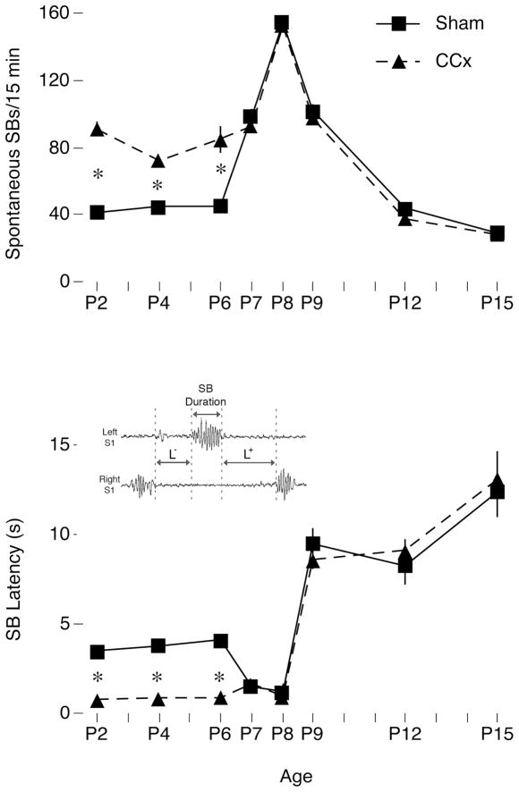
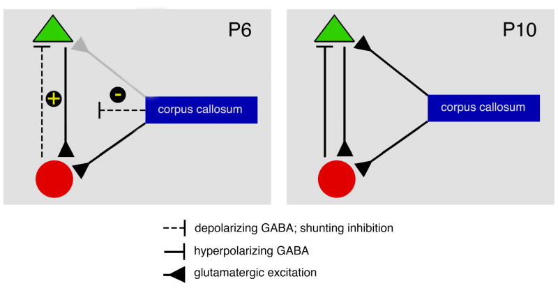
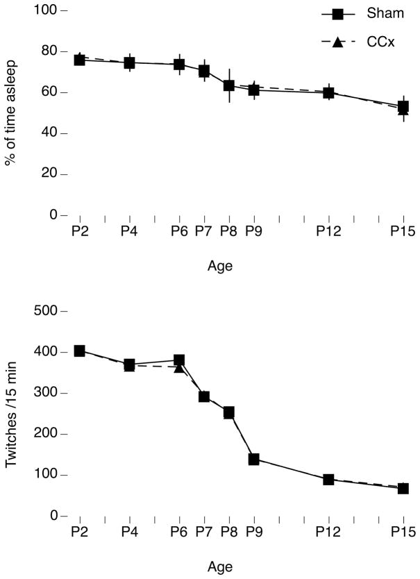
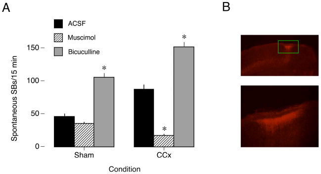
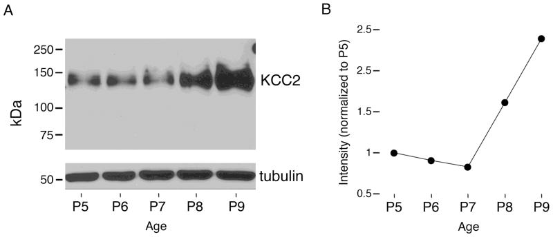
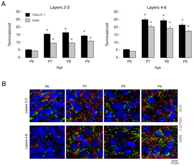
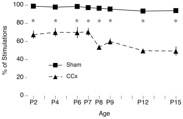
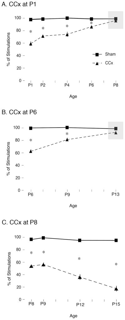
Transecting the corpus callosum of postnatal day (P)1-6 rats disinhibits the production of spindle bursts (SBs) within primary somatosensory cortex (S1), most notably during periods of sleep-related myoclonic twitching. Here we investigated developmental changes in this callosally mediated disinhibition and its association with cortical plasticity. Recordings in P2-15 subjects revealed that callosotomy-induced disinhibition is a transient feature of early development that disappears abruptly after P6. This abrupt switch was accompanied by sharp decreases in myoclonic twitching and equally sharp increases in spontaneous SBs and in the number of GABAergic and glutamatergic presynaptic terminals in S1. Expression of the K+Cl- cotransporter 2 (KCC2) also increased across these ages. To determine whether these developmental changes are associated with alterations in cortical plasticity, pups were callosotomized at P1, P6, or P8, and tested over the subsequent week. Regardless of age, callosotomy immediately disrupted SBs evoked by forepaw stimulation. Over the next week, the P1 and P6 callosotomy groups exhibited full recovery of function; in contrast, the P8 group did not exhibit recovery of function, thus indicating an abrupt decrease in cortical plasticity between P6 and P8. Together, our data demonstrate that callosotomy-induced disinhibition is a transient phenomenon whose disappearance coincides with the onset of increased intrinsic connectivity, establishment of excitatory-inhibitory balance, and diminished plasticity in S1. Accordingly, our findings indicate that callosotomy-induced disinhibition of twitch-related SBs is a bioassay of somatosensory cortical plasticity and, in addition, support the hypothesis that myoclonic twitches, like retinal waves, actively contribute to cortical development and plasticity.
横断出生后第1至6天(P1 - 6)大鼠的胼胝体,会解除初级体感皮层(S1)内纺锤体爆发(SBs)的抑制,在与睡眠相关的肌阵挛抽搐期间尤为明显。在此,我们研究了这种胼胝体介导的去抑制作用的发育变化及其与皮层可塑性的关联。对P2 - 15实验对象的记录显示,胼胝体切开术诱导的去抑制是早期发育的一个短暂特征,在P6之后会突然消失。这种突然转变伴随着肌阵挛抽搐的急剧减少,以及S1中自发性SBs、GABA能和谷氨酸能突触前终末数量的同样急剧增加。钾氯共转运体2(KCC2)的表达在这些年龄段也有所增加。为了确定这些发育变化是否与皮层可塑性的改变有关,在P1、P6或P8对幼崽进行胼胝体切开术,并在随后一周进行测试。无论年龄大小,胼胝体切开术都会立即破坏前爪刺激诱发的SBs。在接下来的一周里,P1和P6胼胝体切开术组表现出功能的完全恢复;相比之下,P8组没有表现出功能恢复,这表明在P6和P8之间皮层可塑性突然下降。总之,我们的数据表明,胼胝体切开术诱导的去抑制是一种短暂现象,其消失与内在连接性增加、兴奋 - 抑制平衡的建立以及S1中可塑性的降低同时发生。因此,我们的研究结果表明,胼胝体切开术诱导的与抽搐相关的SBs的去抑制是体感皮层可塑性的一种生物测定方法,此外,支持了肌阵挛抽搐像视网膜波一样积极促进皮层发育和可塑性的假说。