• 文献检索
  • 文档翻译
  • 深度研究
  • 学术资讯
  • Suppr Zotero 插件Zotero 插件
  • 邀请有礼
  • 套餐&价格
  • 历史记录
应用&插件
Suppr Zotero 插件Zotero 插件浏览器插件Mac 客户端Windows 客户端微信小程序
定价
高级版会员购买积分包购买API积分包
服务
文献检索文档翻译深度研究API 文档MCP 服务
关于我们
关于 Suppr公司介绍联系我们用户协议隐私条款
关注我们

Suppr 超能文献

核心技术专利:CN118964589B侵权必究
粤ICP备2023148730 号-1Suppr @ 2026

文献检索

告别复杂PubMed语法,用中文像聊天一样搜索,搜遍4000万医学文献。AI智能推荐,让科研检索更轻松。

立即免费搜索

文件翻译

保留排版,准确专业,支持PDF/Word/PPT等文件格式,支持 12+语言互译。

免费翻译文档

深度研究

AI帮你快速写综述,25分钟生成高质量综述,智能提取关键信息,辅助科研写作。

立即免费体验

野生型和定位错误的视网膜神经节细胞的体内树突方向发育。

In vivo development of dendritic orientation in wild-type and mislocalized retinal ganglion cells.

机构信息

Department of Biological Structure, University of Washington, Seattle, WA 98195, USA.

出版信息

Neural Dev. 2010 Nov 2;5:29. doi: 10.1186/1749-8104-5-29.

DOI:10.1186/1749-8104-5-29
PMID:21044295
原文链接:https://pmc.ncbi.nlm.nih.gov/articles/PMC2988773/
Abstract

BACKGROUND

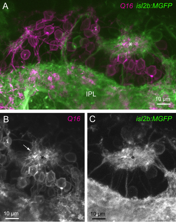
Many neurons in the central nervous system, including retinal ganglion cells (RGCs), possess asymmetric dendritic arbors oriented toward their presynaptic partners. How such dendritic arbors become biased during development in vivo is not well understood. Dendritic arbors may become oriented by directed outgrowth or by reorganization of an initially unbiased arbor. To distinguish between these possibilities, we imaged the dynamic behavior of zebrafish RGC dendrites during development in vivo. We then addressed how cell positioning within the retina, altered in heart-and-soul (has) mutants, affects RGC dendritic orientation.

RESULTS

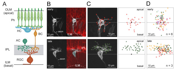
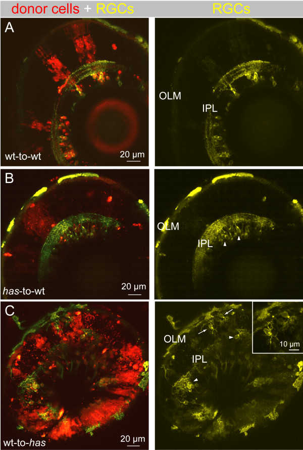
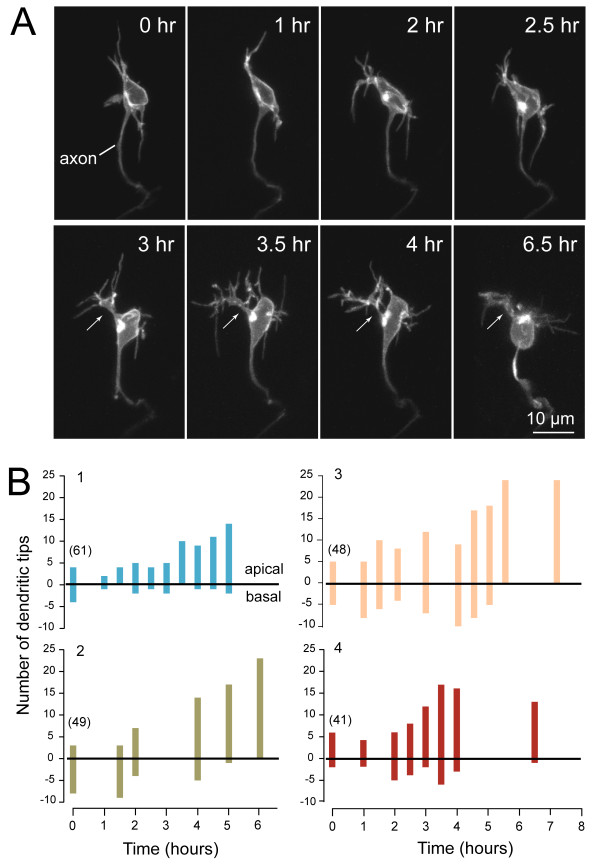
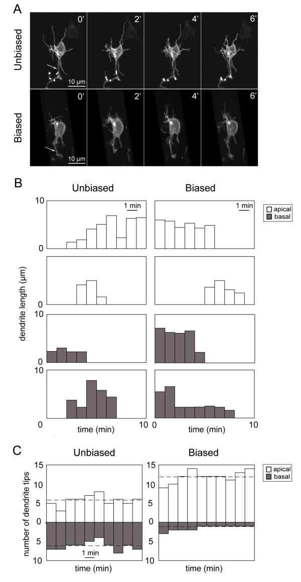
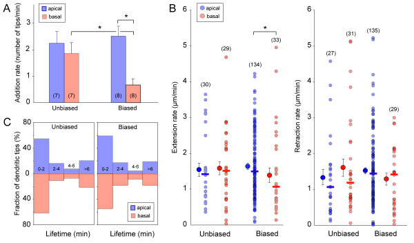
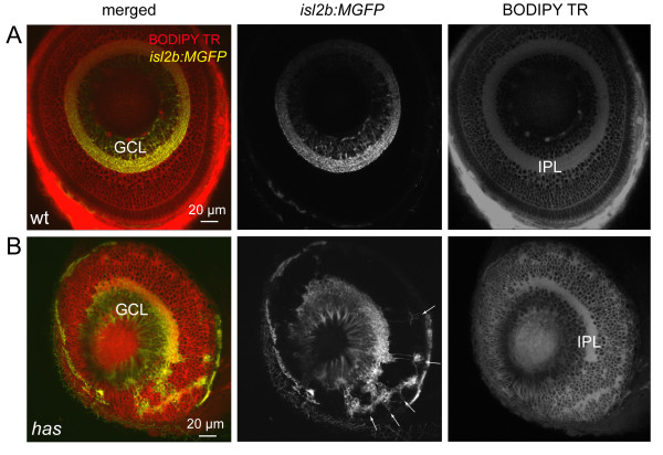
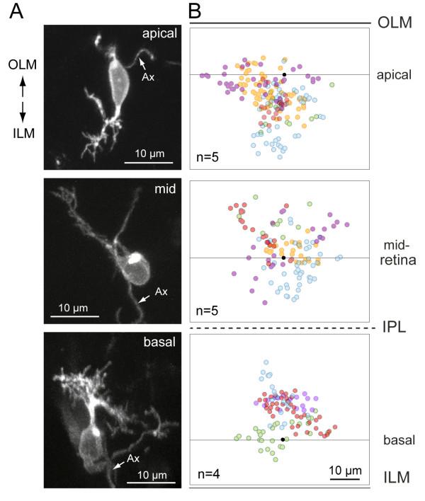
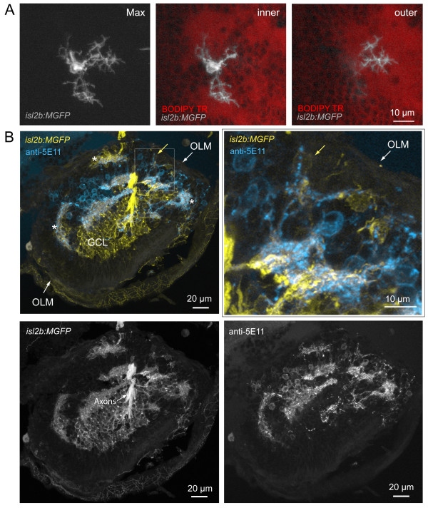
In vivo multiphoton time-lapse analysis revealed that RGC dendrites initially exhibit exploratory behavior in multiple directions but progressively become apically oriented. The lifetimes of basal and apical dendrites were generally comparable before and during the period when arbors became biased. However, with maturation, the addition and extension rates of basal dendrites were slower than those of the apical dendrites. Oriented dendritic arbors were also found in misplaced RGCs of the has retina but there was no preferred orientation amongst the population. However, has RGCs always projected dendrites toward nearby neuropil where amacrine and bipolar cell neurites also terminated. Chimera analysis showed that the abnormal dendritic organization of RGCs in the mutant was non-cell autonomous.

CONCLUSIONS

Our observations show that RGC dendritic arbors acquire an apical orientation by selective and gradual restriction of dendrite addition to the apical side of the cell body, rather than by preferential dendrite stabilization or elimination. A biased arbor emerges at a stage when many of the dendritic processes still appear exploratory. The generation of an oriented RGC dendritic arbor is likely to be determined by cell-extrinsic cues. Such cues are unlikely to be localized to the basal lamina of the inner retina, but rather may be provided by cells presynaptic to the RGCs.

摘要

背景

中枢神经系统中的许多神经元,包括视网膜神经节细胞(RGCs),具有朝向其突触前伙伴的不对称树突分支。在体内发育过程中,这些树突分支如何变得偏向尚不清楚。树突分支可能通过定向生长或最初无偏树突的重组来定向。为了区分这些可能性,我们在体内成像了斑马鱼 RGC 树突在发育过程中的动态行为。然后,我们研究了视网膜内细胞定位的变化如何影响 RGC 树突的方向,这种变化在心脏与灵魂(has)突变体中发生。

结果

体内多光子延时分析显示,RGC 树突最初表现出多方向的探索行为,但逐渐变得向顶端定向。在树突变得偏向之前和期间,基底和顶端树突的寿命通常相似。然而,随着成熟,基底树突的添加和延伸速度比顶端树突慢。在 has 视网膜中错位的 RGC 中也发现了定向树突,但在群体中没有优先方向。然而,has RGC 总是将树突投射到附近的神经胶质中,那里也终止了无长突细胞和双极细胞的轴突。嵌合体分析表明,突变体中 RGC 的异常树突组织是非细胞自主的。

结论

我们的观察表明,RGC 树突通过选择性和逐渐限制树突向细胞体顶端的添加来获得顶端取向,而不是通过优先稳定或消除树突。在许多树突过程仍然表现出探索性的阶段,出现了偏向的树突。有向 RGC 树突的产生可能取决于细胞外的线索。这些线索不太可能局限于内视网膜的基底层,而可能是由 RGC 突触前的细胞提供的。

https://cdn.ncbi.nlm.nih.gov/pmc/blobs/fd31/2988773/b7c3998b44ba/1749-8104-5-29-9.jpg
https://cdn.ncbi.nlm.nih.gov/pmc/blobs/fd31/2988773/47da476c23d4/1749-8104-5-29-1.jpg
https://cdn.ncbi.nlm.nih.gov/pmc/blobs/fd31/2988773/a61284d59e86/1749-8104-5-29-2.jpg
https://cdn.ncbi.nlm.nih.gov/pmc/blobs/fd31/2988773/4a5762bfc3b6/1749-8104-5-29-3.jpg
https://cdn.ncbi.nlm.nih.gov/pmc/blobs/fd31/2988773/b571e258139e/1749-8104-5-29-4.jpg
https://cdn.ncbi.nlm.nih.gov/pmc/blobs/fd31/2988773/78f65c23f317/1749-8104-5-29-5.jpg
https://cdn.ncbi.nlm.nih.gov/pmc/blobs/fd31/2988773/a84813805945/1749-8104-5-29-6.jpg
https://cdn.ncbi.nlm.nih.gov/pmc/blobs/fd31/2988773/7556a94b5648/1749-8104-5-29-7.jpg
https://cdn.ncbi.nlm.nih.gov/pmc/blobs/fd31/2988773/06e47651b5e7/1749-8104-5-29-8.jpg
https://cdn.ncbi.nlm.nih.gov/pmc/blobs/fd31/2988773/b7c3998b44ba/1749-8104-5-29-9.jpg

相似文献

1
In vivo development of dendritic orientation in wild-type and mislocalized retinal ganglion cells.野生型和定位错误的视网膜神经节细胞的体内树突方向发育。
Neural Dev. 2010 Nov 2;5:29. doi: 10.1186/1749-8104-5-29.
2
In vivo imaging reveals dendritic targeting of laminated afferents by zebrafish retinal ganglion cells.体内成像揭示斑马鱼视网膜神经节细胞对分层传入纤维的树突靶向作用。
Neuron. 2006 Nov 22;52(4):609-21. doi: 10.1016/j.neuron.2006.10.004.
3
Cellular and Molecular Analysis of Dendritic Morphogenesis in a Retinal Cell Type That Senses Color Contrast and Ventral Motion.对一种感知颜色对比度和腹侧运动的视网膜细胞类型中树突形态发生的细胞和分子分析。
J Neurosci. 2017 Dec 13;37(50):12247-12262. doi: 10.1523/JNEUROSCI.2098-17.2017. Epub 2017 Nov 7.
4
Factors controlling the dendritic arborization of retinal ganglion cells.控制视网膜神经节细胞树突分支的因素。
Vis Neurosci. 1996 Jul-Aug;13(4):721-33. doi: 10.1017/s0952523800008609.
5
Local and target-derived brain-derived neurotrophic factor exert opposing effects on the dendritic arborization of retinal ganglion cells in vivo.局部和靶源性脑源性神经营养因子对体内视网膜神经节细胞的树突分支具有相反的作用。
J Neurosci. 2002 Sep 1;22(17):7639-49. doi: 10.1523/JNEUROSCI.22-17-07639.2002.
6
A general principle governs vision-dependent dendritic patterning of retinal ganglion cells.一条普遍原则支配着视网膜神经节细胞依赖视觉的树突形态形成。
J Comp Neurol. 2014 Oct 15;522(15):3403-22. doi: 10.1002/cne.23609. Epub 2014 Apr 29.
7
Developmental mechanisms that regulate retinal ganglion cell dendritic morphology.调控视网膜神经节细胞树突形态发生的发育机制。
Dev Neurobiol. 2011 Dec;71(12):1297-309. doi: 10.1002/dneu.20900.
8
Cell-autonomous TrkB signaling in presynaptic retinal ganglion cells mediates axon arbor growth and synapse maturation during the establishment of retinotectal synaptic connectivity.视网膜神经节细胞突触前的细胞自主TrkB信号传导在视网膜-脑顶盖突触连接建立过程中介导轴突分支生长和突触成熟。
J Neurosci. 2007 Mar 7;27(10):2444-56. doi: 10.1523/JNEUROSCI.4434-06.2007.
9
DSCAM differentially modulates pre- and postsynaptic structural and functional central connectivity during visual system wiring.DSCAM 差异调节视觉系统布线过程中突触前和突触后结构和功能的中枢连接。
Neural Dev. 2018 Sep 15;13(1):22. doi: 10.1186/s13064-018-0118-5.
10
Strip1 regulates retinal ganglion cell survival by suppressing Jun-mediated apoptosis to promote retinal neural circuit formation.Strip1 通过抑制 Jun 介导的细胞凋亡来调节视网膜神经节细胞的存活,从而促进视网膜神经回路的形成。
Elife. 2022 Mar 22;11:e74650. doi: 10.7554/eLife.74650.

引用本文的文献

1
Prevalence and practices of immunofluorescent cell image processing: a systematic review.免疫荧光细胞图像处理的患病率与实践:一项系统综述
Front Cell Neurosci. 2023 Jul 20;17:1188858. doi: 10.3389/fncel.2023.1188858. eCollection 2023.
2
Strip1 regulates retinal ganglion cell survival by suppressing Jun-mediated apoptosis to promote retinal neural circuit formation.Strip1 通过抑制 Jun 介导的细胞凋亡来调节视网膜神经节细胞的存活,从而促进视网膜神经回路的形成。
Elife. 2022 Mar 22;11:e74650. doi: 10.7554/eLife.74650.
3
Visual Experience Influences Dendritic Orientation but Is Not Required for Asymmetric Wiring of the Retinal Direction Selective Circuit.

本文引用的文献

1
Analysis of aPKClambda and aPKCzeta reveals multiple and redundant functions during vertebrate retinogenesis.对非典型蛋白激酶Cλ(aPKClambda)和非典型蛋白激酶Cζ(aPKCzeta)的分析揭示了脊椎动物视网膜发育过程中的多种冗余功能。
Mol Cell Neurosci. 2007 Mar;34(3):431-44. doi: 10.1016/j.mcn.2006.11.016. Epub 2007 Jan 12.
2
Polarization and orientation of retinal ganglion cells in vivo.视网膜神经节细胞在体内的极化与取向
Neural Dev. 2006 Oct 13;1:2. doi: 10.1186/1749-8104-1-2.
3
In vivo imaging reveals dendritic targeting of laminated afferents by zebrafish retinal ganglion cells.
视觉体验影响树突的方向,但不是视网膜方向选择回路不对称布线所必需的。
Cell Rep. 2020 Jun 30;31(13):107844. doi: 10.1016/j.celrep.2020.107844.
4
Diagnostic Ability of Macular Vessel Density in the Ganglion Cell-Inner Plexiform Layer on Optical Coherence Tomographic Angiography for Glaucoma.光学相干断层扫描血管造影术测量神经节细胞-内层神经纤维层黄斑血管密度对青光眼的诊断能力
Transl Vis Sci Technol. 2019 Jul 30;8(4):12. doi: 10.1167/tvst.8.4.12. eCollection 2019 Jul.
5
Let's get small (and smaller): Combining zebrafish and nanomedicine to advance neuroregenerative therapeutics.让我们变得更小(更小):结合斑马鱼和纳米医学推进神经再生治疗。
Adv Drug Deliv Rev. 2019 Aug;148:344-359. doi: 10.1016/j.addr.2019.01.011. Epub 2019 Feb 12.
6
An Antagonistic Axon-Dendrite Interplay Enables Efficient Neuronal Repair in the Adult Zebrafish Central Nervous System.拮抗轴突-树突相互作用使成年斑马鱼中枢神经系统的神经元修复更有效。
Mol Neurobiol. 2019 May;56(5):3175-3192. doi: 10.1007/s12035-018-1292-5. Epub 2018 Aug 13.
7
Opportunities and challenges for using the zebrafish to study neuronal connectivity as an endpoint of developmental neurotoxicity.利用斑马鱼研究发育神经毒性的神经元连接作为终点的机遇与挑战。
Neurotoxicology. 2018 Jul;67:102-111. doi: 10.1016/j.neuro.2018.04.016. Epub 2018 Apr 25.
8
Segmented inner plexiform layer thickness as a potential biomarker to evaluate open-angle glaucoma: Dendritic degeneration of retinal ganglion cell.分段内网状层厚度作为评估开角型青光眼的潜在生物标志物:视网膜神经节细胞的树突状变性
PLoS One. 2017 Aug 3;12(8):e0182404. doi: 10.1371/journal.pone.0182404. eCollection 2017.
9
Mechanisms regulating dendritic arbor patterning.调节树突状分支模式的机制。
Cell Mol Life Sci. 2017 Dec;74(24):4511-4537. doi: 10.1007/s00018-017-2588-8. Epub 2017 Jul 22.
10
Imaging the adult zebrafish cone mosaic using optical coherence tomography.使用光学相干断层扫描对成年斑马鱼视锥细胞镶嵌结构进行成像。
Vis Neurosci. 2016 Jan;33:E011. doi: 10.1017/S0952523816000092.
体内成像揭示斑马鱼视网膜神经节细胞对分层传入纤维的树突靶向作用。
Neuron. 2006 Nov 22;52(4):609-21. doi: 10.1016/j.neuron.2006.10.004.
4
In vivo development of retinal ON-bipolar cell axonal terminals visualized in nyx::MYFP transgenic zebrafish.在nyx::MYFP转基因斑马鱼中可视化的视网膜ON双极细胞轴突终末的体内发育。
Vis Neurosci. 2006 Sep-Oct;23(5):833-43. doi: 10.1017/S0952523806230219.
5
Time frame of mitral cell development in the mice olfactory bulb.小鼠嗅球中二尖瓣细胞的发育时间框架。
J Comp Neurol. 2006 Jun 1;496(4):529-43. doi: 10.1002/cne.20941.
6
The level and integrity of synaptic input regulates dendrite structure.突触输入的水平和完整性调节树突结构。
J Neurosci. 2006 Feb 1;26(5):1539-50. doi: 10.1523/JNEUROSCI.3807-05.2006.
7
Polarized secretory trafficking directs cargo for asymmetric dendrite growth and morphogenesis.极化分泌运输引导货物进行不对称树突生长和形态发生。
Neuron. 2005 Dec 8;48(5):757-71. doi: 10.1016/j.neuron.2005.11.005.
8
Targeting of amacrine cell neurites to appropriate synaptic laminae in the developing zebrafish retina.在发育中的斑马鱼视网膜中,无长突细胞神经突靶向至适当的突触层。
Development. 2005 Nov;132(22):5069-79. doi: 10.1242/dev.02075.
9
Physiological and morphological development of the rat cerebellar Purkinje cell.大鼠小脑浦肯野细胞的生理和形态发育
J Physiol. 2005 Sep 15;567(Pt 3):829-50. doi: 10.1113/jphysiol.2005.089383. Epub 2005 Jul 7.
10
A GFP-based genetic screen reveals mutations that disrupt the architecture of the zebrafish retinotectal projection.一项基于绿色荧光蛋白的遗传筛选揭示了破坏斑马鱼视网膜顶盖投射结构的突变。
Development. 2005 Jul;132(13):2955-67. doi: 10.1242/dev.01861. Epub 2005 Jun 1.