Xu Shunjiang, Flanagan Judith L, Simmons Peter A, Vehige Joseph, Willcox Mark D, Garrett Qian
Brien Holden Vision Institute, The University of New South Wales, Sydney, NSW, Australia.
Mol Vis. 2010 Sep 4;16:1823-31.
Previously we demonstrated expression and localization of carnitine/organic cation transporters, OCTN1 and OCTN2, in human corneal and conjunctival epithelia. The present study aimed to examine the characteristics of L-carnitine transporters in cultured human limbal corneal (HCLE) and conjunctival epithelial (HCjE) cells.
Time-course, Na(+)-dependence, kinetics, energy- and pH- dependence of L-carnitine transport were investigated by monitoring L-[(3)H]carnitine uptake into HCLE and HCjE cells. To determine the specificity of action, competition and inhibition studies were performed.
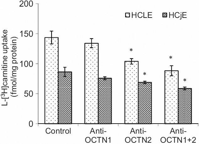
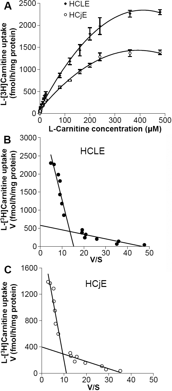
The uptake of L-carnitine into HCLE and HCjE cells was saturable and time-dependent. An Eadie-Hofstee plot showed two distinct components: a high- and a low- affinity carnitine transport system in HCLE and/or HCjE cells. L-carnitine transport was significantly inhibited by the metabolic inhibitors (sodium azide, dinitrophenol, iodoacetic acid). The L-carnitine analogs (D-carnitine, acetyl-L-carnitine and γ-butyrobetaine), tetraethylammonium (TEA), 2-amino-2-norbornane carboxylic acid (BCH), strongly inhibited uptake of L-[(3)H]carnitine. Uptake of L-[(3)H]carnitine also required the presence of Na(+) in the external medium and the uptake activity was maximal at pH 5.5. The anti-OCTN2 antibody blocked L-carnitine uptake in both HCLE and HCjE cells whereas the anti-OCTN1 antibody did not significantly block L-carnitine uptake.
L-carnitine is transported into HCLE and HCjE cells by an active carrier mediated transport system that is time-, Na(+)-, energy- and pH- dependent. The carnitine/organic cation transporter OCTN2 appears to play a dominant role in this process.
此前我们已证明肉碱/有机阳离子转运体OCTN1和OCTN2在人角膜和结膜上皮中的表达及定位。本研究旨在检测培养的人角膜缘上皮(HCLE)细胞和结膜上皮(HCjE)细胞中左旋肉碱转运体的特征。
通过监测L-[(3)H]肉碱进入HCLE和HCjE细胞的摄取情况,研究左旋肉碱转运的时间进程、对钠离子的依赖性、动力学、能量和pH依赖性。为确定作用的特异性,进行了竞争和抑制研究。
L-肉碱进入HCLE和HCjE细胞的摄取是可饱和的且具有时间依赖性。伊迪-霍夫斯蒂图显示有两个不同的成分:HCLE和/或HCjE细胞中一个高亲和力和一个低亲和力的肉碱转运系统。代谢抑制剂(叠氮化钠、二硝基苯酚、碘乙酸)显著抑制L-肉碱转运。L-肉碱类似物(D-肉碱、乙酰-L-肉碱和γ-丁基甜菜碱)、四乙铵(TEA)、2-氨基-2-降冰片烷羧酸(BCH)强烈抑制L-[(3)H]肉碱的摄取。L-[(3)H]肉碱的摄取还需要细胞外培养基中存在钠离子,且摄取活性在pH 5.5时最大。抗OCTN2抗体可阻断HCLE和HCjE细胞中L-肉碱的摄取,而抗OCTN1抗体未显著阻断L-肉碱的摄取。
L-肉碱通过一个依赖时间、钠离子、能量和pH的主动载体介导转运系统进入HCLE和HCjE细胞。肉碱/有机阳离子转运体OCTN2似乎在此过程中起主导作用。