Rose David Z, Parra-Herran Carlos, Petito Carol K, Post M Judith D
Department of Neurology, University of Miami Miller School of Medicine, Jackson Memorial Hospital, Miami, Fla., USA.
Case Rep Neurol. 2010 Aug 7;2(2):101-110. doi: 10.1159/000319691.
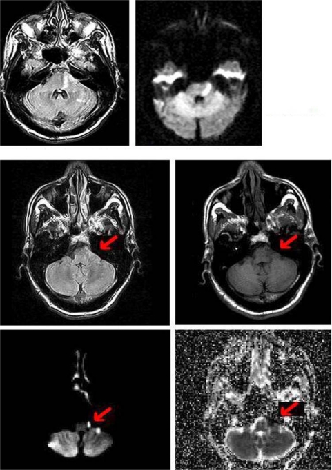
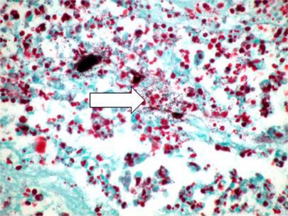
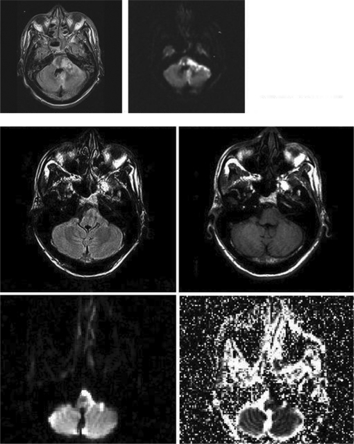
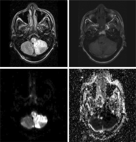
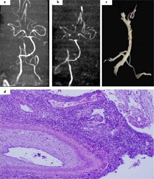
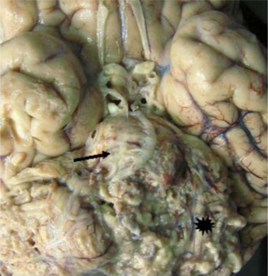
Extra-axial restriction on diffusion weighted imaging (DWI) is an unusual finding on brain magnetic resonance imaging (MRI). Intra-axial restriction on DWI, however, is common, and can represent brain parenchymal infarction, tumor, abscess, or toxic-metabolic process. The infrequency of extra-axial DWI restriction and the paucity of clinico-pathological correlation in the literature limit its differential diagnosis. Scant case reports suggest that extra-axial DWI restriction could be a lymphoma, neurenteric cyst, or, in one patient, subdural empyema [1,2,3]. We postulate that pus formation must be excluded first, because it can provoke an aggressive meningo-vasculitis with rapidly fatal, intra-axial infarctions. Our patient was a 45-year-old man, presenting to our hospital with left facial droop and right (contralateral) arm and leg weakness. Initial MRI revealed DWI restriction in the left lateral pons, consistent with a classic Millard-Gubler stroke. Also noted was a subtle, extra-axial area of curvilinear diffusion restriction in the left cerebellar-pontine angle's subarachnoid space. Days later, the patient had a headache, and repeat MRI revealed extension of the two DWI lesions - both the intra-axial pontine infarction and the extra-axial area of restricted diffusion in the subarachnoid space. The patient became comatose, a third MRI revealed more extensive DWI restrictions, and he expired despite aggressive care. Autopsy revealed massive brainstem infarcts, a thick lymphoplasmacytic infiltrate, copious Gram-Positive cocci (likely MRSA) and arteries partially occluded with fibrointimal proliferation. This emphasizes the concept that extra-axial DWI restriction can represent pus development in the subarachnoid space - a radiographic marker to identify a patient at risk for demise due to septic, meningo-vasculitic infarctions.
扩散加权成像(DWI)上的轴外扩散受限是脑磁共振成像(MRI)上一种不常见的表现。然而,DWI上的轴内扩散受限很常见,可提示脑实质梗死、肿瘤、脓肿或中毒代谢过程。轴外DWI受限情况少见,且文献中临床病理相关性资料匮乏,限制了其鉴别诊断。少量病例报告提示轴外DWI受限可能为淋巴瘤、神经肠囊肿,或在1例患者中为硬膜下积脓[1,2,3]。我们推测必须首先排除脓液形成,因为其可引发侵袭性脑膜血管炎,导致迅速致命的轴内梗死。我们的患者为一名45岁男性,因左侧面部下垂及右侧(对侧)手臂和腿部无力入院。初始MRI显示左侧脑桥外侧DWI受限,符合典型的Millard - Gubler卒中表现。同时还注意到左侧小脑脑桥角蛛网膜下腔内有一个细微的、呈曲线状的扩散受限轴外区域。数日后,患者出现头痛,复查MRI显示两个DWI病灶均有扩大——轴内脑桥梗死及蛛网膜下腔内扩散受限的轴外区域。患者陷入昏迷,第三次MRI显示更广泛的DWI受限,尽管积极治疗仍死亡。尸检发现大量脑干梗死、厚层淋巴浆细胞浸润、大量革兰氏阳性球菌(可能为耐甲氧西林金黄色葡萄球菌)以及部分被纤维内膜增生阻塞的动脉。这强调了轴外DWI受限可提示蛛网膜下腔内脓液形成这一概念——这是一种影像学标志物,可用于识别因感染性脑膜血管炎梗死而有死亡风险的患者。