Cho Yang Kyeung, Chang Hwa Seok, La Tae Yoon, Ji Donghyun, Kim Hyunkyung, Choi Jin A, Kim Man Soo
Department of Ophthalmology, St. Vincent's Hospital, The Catholic University of Korea School of Medicine, Suwon, Korea.
Korean J Ophthalmol. 2010 Oct;24(5):284-90. doi: 10.3341/kjo.2010.24.5.284. Epub 2010 Oct 5.
We evaluated various preoperative anterior segment parameters measured with a Pentacam rotating Scheimpflug camera and compared them with those of conventional methods. We also evaluated the effect of different parameters on corneal endothelial cells after cataract surgery.
Pentacam examination was performed in 88 eyes from 88 patients to evaluate central anterior chamber depth (ACD(pentacam)), nuclear density (Densitometry(pentacam)), anterior chamber volume (ACV), and lens thickness (LT(pentacam)). We compared values of ACD(pentacam) with those of ultrasound (ACD(sono)) and also compared Densitometry(pentacam) values with those of Lens Opacities Classification System (LOCS III) classification. We evaluated the effect of the following preoperative values measured with Pentacam on postoperative endothelial cell loss: pupil size measured both preoperatively and before capsulorrhexsis (Pupil(CCC)), amount of viscoelastics, and LT measured by ultrasound (LT(sono)).
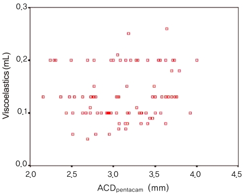
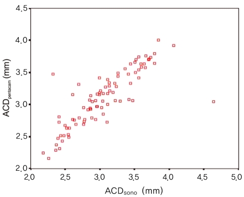
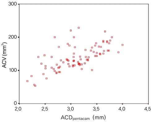
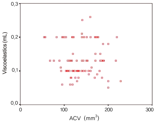
A SIGNIFICANT CONCORDANCE WAS FOUND BETWEEN THE TWO GRADING METHODS OF NUCLEAR OPACITY: Densitometry(pentacam) and LOCS III classification (τ(b) = 0.414, p = 0.000). We also found a positive correlation between ACD(pentacam) and ACD(sono) (r = 0.823, p = 0.000) and between ACD(pentacam) and ACV (r = 0.650, p = 0.000). There were significant differences between the results of LT(pentacam) and LT(sono). The final regression model identified Densitometry(pentacam), viscoelastics and Pupil(CCC) as independent predictors of decreased postoperative corneal endothelial cell density (CD) at postoperative day 3, and Densitometry(pentacam), viscoelastics, and ACV as independent predictors of decreased CD two months postoperatively (p<0.05).
Good agreement was found between all results obtained with the Pentacam and conventional methods except LT. Analyzing anterior chamber parameters preoperatively using Pentacam could be helpful to predict postoperative endothelial cell loss.
我们评估了使用Pentacam旋转式Scheimpflug相机测量的各种术前眼前节参数,并将其与传统方法测量的参数进行比较。我们还评估了白内障手术后不同参数对角膜内皮细胞的影响。
对88例患者的88只眼进行Pentacam检查,以评估中央前房深度(ACD(Pentacam))、核密度(密度测量法(Pentacam))、前房容积(ACV)和晶状体厚度(LT(Pentacam))。我们将ACD(Pentacam)的值与超声测量的值(ACD(超声))进行比较,并将密度测量法(Pentacam)的值与晶状体混浊分类系统(LOCS III)分类的值进行比较。我们评估了Pentacam术前测量的以下值对术后内皮细胞损失的影响:术前和撕囊前测量的瞳孔大小(瞳孔(CCC))、粘弹剂用量以及超声测量的LT(LT(超声))。
在核混浊的两种分级方法之间发现了显著的一致性:密度测量法(Pentacam)和LOCS III分类(τ(b)=0.414,p=0.000)。我们还发现ACD(Pentacam)与ACD(超声)之间呈正相关(r=0.823,p=0.000),ACD(Pentacam)与ACV之间呈正相关(r=0.650,p=0.000)。LT(Pentacam)和LT(超声)的结果存在显著差异。最终回归模型确定密度测量法(Pentacam)、粘弹剂和瞳孔(CCC)是术后第3天角膜内皮细胞密度(CD)降低的独立预测因素,密度测量法(Pentacam)、粘弹剂和ACV是术后两个月CD降低的独立预测因素(p<0.05)。
除LT外,Pentacam获得的所有结果与传统方法之间均发现良好的一致性。术前使用Pentacam分析前房参数可能有助于预测术后内皮细胞损失。