Cardiovascular Research Institute, University of California San Francisco, San Francisco, California, United States of America.
PLoS One. 2010 Oct 29;5(10):e13689. doi: 10.1371/journal.pone.0013689.
The notochord is a signaling center required for the patterning of the vertebrate embryonic midline, however, the molecular and cellular mechanisms involved in the formation of this essential embryonic tissue remain unclear. The urochordate Ciona intestinalis develops a simple notochord from 40 specific postmitotic mesodermal cells. The precursors intercalate mediolaterally and establish a single array of disk-shaped notochord cells along the midline. However, the role that notochord precursor polarization, particularly along the dorsoventral axis, plays in this morphogenetic process remains poorly understood.
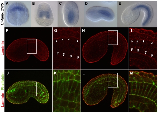
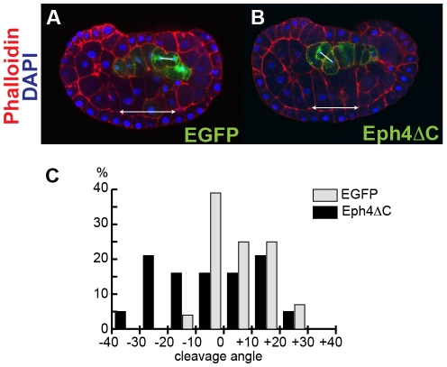
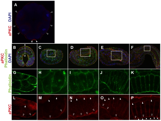
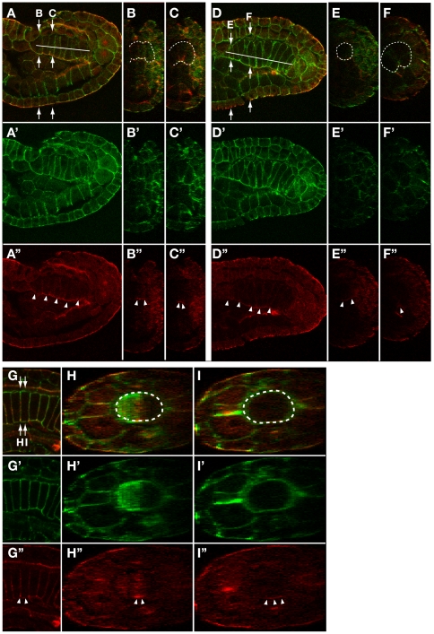
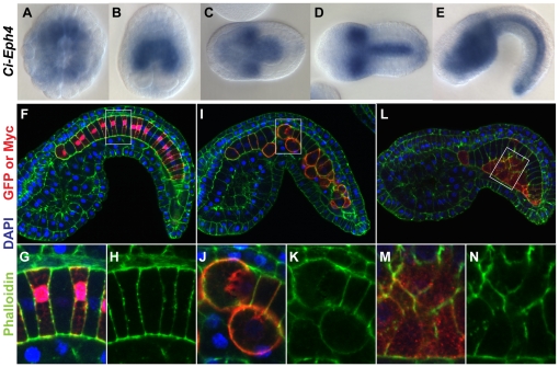
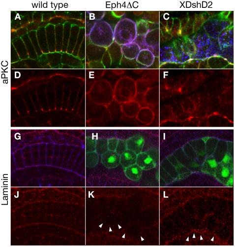
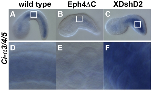
METHODOLOGY/PRINCIPAL FINDINGS: Here we show that the notochord preferentially accumulates an apical cell polarity marker, aPKC, ventrally and a basement membrane marker, laminin, dorsally. This asymmetric accumulation of apicobasal cell polarity markers along the embryonic dorsoventral axis was sustained in notochord precursors during convergence and extension. Further, of several members of the Eph gene family implicated in cellular and tissue morphogenesis, only Ci-Eph4 was predominantly expressed in the notochord throughout cell intercalation. Introduction of a dominant-negative Ci-Eph4 to notochord precursors diminished asymmetric accumulation of apicobasal cell polarity markers, leading to defective intercalation. In contrast, misexpression of a dominant-negative mutant of a planar cell polarity gene Dishevelled preserved asymmetric accumulation of aPKC and laminin in notochord precursors, although their intercalation was incomplete.
CONCLUSIONS/SIGNIFICANCE: Our data support a model in which in ascidian embryos Eph-dependent dorsoventral polarity of notochord precursors plays a crucial role in mediolateral cell intercalation and is required for proper notochord morphogenesis.
脊索是脊椎动物胚胎中中线模式形成所必需的信号中心,然而,形成这种重要胚胎组织的分子和细胞机制仍不清楚。尾索动物文昌鱼由 40 个特定的有丝分裂后中胚层细胞发育出简单的脊索。前体细胞在中侧方向上插入,并在中线建立一个单一的盘状脊索细胞阵列。然而,脊索前体细胞极化的作用,特别是沿着背腹轴的极化,在这个形态发生过程中仍然知之甚少。
方法/主要发现:在这里,我们发现脊索优先在腹侧积累顶端细胞极性标记物 aPKC,在背侧积累基底膜标记物 laminin。这种沿着胚胎背腹轴的顶端-基底细胞极性标记物的不对称积累在脊索前体细胞的会聚和延伸过程中得以维持。此外,在几个参与细胞和组织形态发生的 Eph 基因家族成员中,只有 Ci-Eph4 在细胞插入过程中主要在脊索中表达。向脊索前体细胞中引入显性负性的 Ci-Eph4 会减少顶端-基底细胞极性标记物的不对称积累,导致插入缺陷。相比之下,Dishevelled 的显性负突变体的异常表达虽然不完全,但在脊索前体细胞中保持了 aPKC 和 laminin 的不对称积累。
结论/意义:我们的数据支持这样一种模型,即在文昌鱼胚胎中 Eph 依赖性脊索前体细胞的背腹极性在介导中侧方向的细胞插入中起着至关重要的作用,并且是脊索正常形态发生所必需的。