Laboratorio de Inmunología y Virología, Departamento de Microbiología e Inmunología, Facultad de Ciencias Biológicas de la Universidad Autónoma de Nuevo León, San Nicolás de los Garza, N, L, México.
J Exp Clin Cancer Res. 2010 Nov 16;29(1):148. doi: 10.1186/1756-9966-29-148.
Colloidal silver has been used as an antimicrobial and disinfectant agent. However, there is scarce information on its antitumor potential. The aim of this study was to determine if colloidal silver had cytotoxic effects on MCF-7 breast cancer cells and its mechanism of cell death.
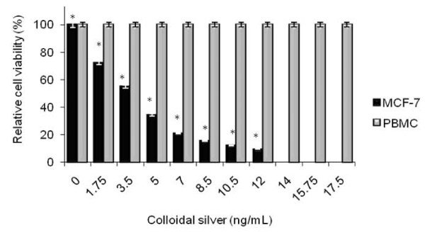
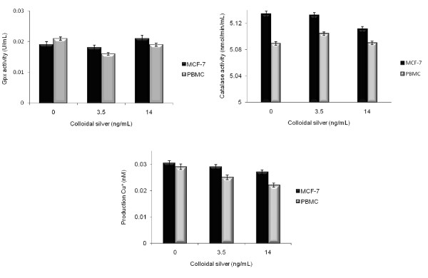
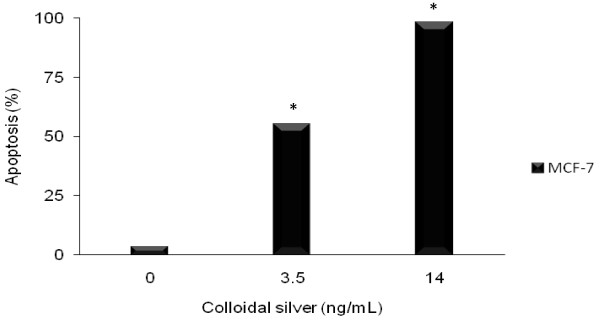
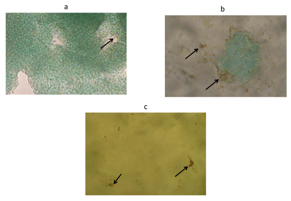
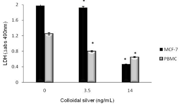
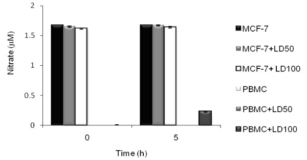
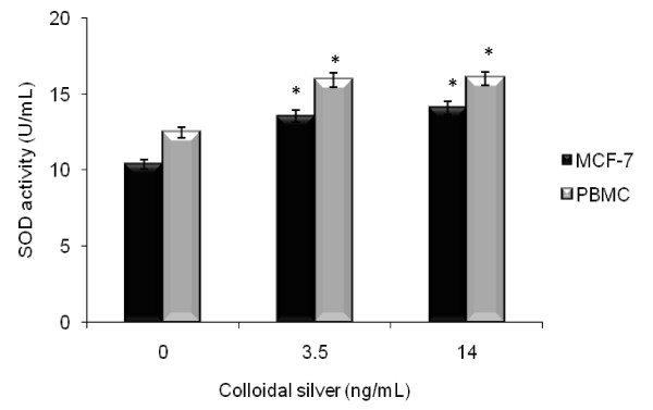
MCF-7 breast cancer cells were treated with colloidal silver (ranged from 1.75 to 17.5 ng/mL) for 5 h at 37°C and 5% CO2 atmosphere. Cell Viability was evaluated by trypan blue exclusion method and the mechanism of cell death through detection of mono-oligonucleosomes using an ELISA kit and TUNEL assay. The production of NO, LDH, and Gpx, SOD, CAT, and Total antioxidant activities were evaluated by colorimetric assays.
Colloidal silver had dose-dependent cytotoxic effect in MCF-7 breast cancer cells through induction of apoptosis, shown an LD50 (3.5 ng/mL) and LD100 (14 ng/mL) (*P < 0.05), significantly decreased LDH (*P < 0.05) and significantly increased SOD (*P < 0.05) activities. However, the NO production, and Gpx, CAT, and Total antioxidant activities were not affected in MCF-7 breast cancer cells. PBMC were not altered by colloidal silver.
The present results showed that colloidal silver might be a potential alternative agent for human breast cancer therapy.
胶体银已被用作一种抗菌和消毒剂。然而,关于其抗肿瘤潜力的信息却很少。本研究的目的是确定胶体银对 MCF-7 乳腺癌细胞是否具有细胞毒性作用及其细胞死亡的机制。
MCF-7 乳腺癌细胞在 37°C 和 5%CO2 环境下用胶体银(浓度范围为 1.75 至 17.5ng/ml)处理 5 小时。用台盼蓝排除法评估细胞活力,并通过 ELISA 试剂盒和 TUNEL 检测检测单聚体寡核苷酸来检测细胞死亡的机制。通过比色法评估 NO、LDH、Gpx、SOD、CAT 和总抗氧化活性的产生。
胶体银对 MCF-7 乳腺癌细胞具有剂量依赖性的细胞毒性作用,通过诱导细胞凋亡来实现,表现出 LD50(3.5ng/ml)和 LD100(14ng/ml)(*P<0.05),显著降低 LDH(*P<0.05)并显著增加 SOD(*P<0.05)活性。然而,NO 产生、Gpx、CAT 和总抗氧化活性在 MCF-7 乳腺癌细胞中没有受到影响。胶体银对 PBMC 没有改变。
本研究结果表明,胶体银可能是人类乳腺癌治疗的一种潜在替代药物。