Laboratory for Molecular Neurogenesis, RIKEN Brain Science Institute, Wako, Saitama, Japan.
PLoS One. 2010 Nov 11;5(11):e13932. doi: 10.1371/journal.pone.0013932.
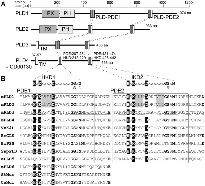
Phospholipase D (PLD) catalyzes conversion of phosphatidylcholine into choline and phosphatidic acid, leading to a variety of intracellular signal transduction events. Two classical PLDs, PLD1 and PLD2, contain phosphatidylinositide-binding PX and PH domains and two conserved His-x-Lys-(x)(4)-Asp (HKD) motifs, which are critical for PLD activity. PLD4 officially belongs to the PLD family, because it possesses two HKD motifs. However, it lacks PX and PH domains and has a putative transmembrane domain instead. Nevertheless, little is known regarding expression, structure, and function of PLD4.
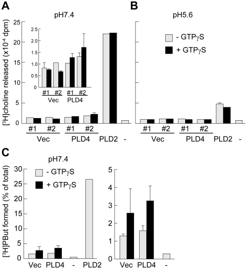
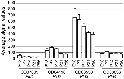
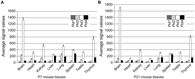
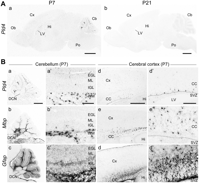
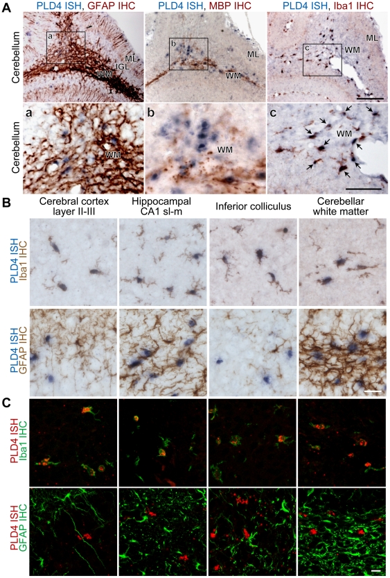
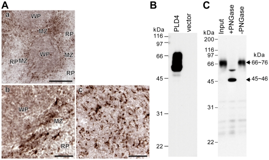
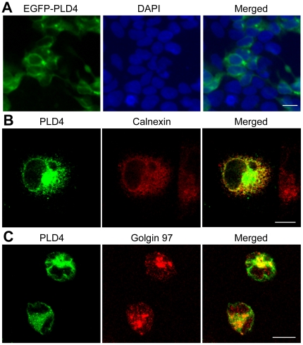
METHODOLOGY/PRINCIPAL FINDINGS: PLD4 was analyzed in terms of expression, structure, and function. Expression was analyzed in developing mouse brains and non-neuronal tissues using microarray, in situ hybridization, immunohistochemistry, and immunocytochemistry. Structure was evaluated using bioinformatics analysis of protein domains, biochemical analyses of transmembrane property, and enzymatic deglycosylation. PLD activity was examined by choline release and transphosphatidylation assays. Results demonstrated low to modest, but characteristic, PLD4 mRNA expression in a subset of cells preferentially localized around white matter regions, including the corpus callosum and cerebellar white matter, during the first postnatal week. These PLD4 mRNA-expressing cells were identified as Iba1-positive microglia. In non-neuronal tissues, PLD4 mRNA expression was widespread, but predominantly distributed in the spleen. Intense PLD4 expression was detected around the marginal zone of the splenic red pulp, and splenic PLD4 protein recovered from subcellular membrane fractions was highly N-glycosylated. PLD4 was heterologously expressed in cell lines and localized in the endoplasmic reticulum and Golgi apparatus. Moreover, heterologously expressed PLD4 proteins did not exhibit PLD enzymatic activity.
CONCLUSIONS/SIGNIFICANCE: Results showed that PLD4 is a non-PLD, HKD motif-carrying, transmembrane glycoprotein localized in the endoplasmic reticulum and Golgi apparatus. The spatiotemporally restricted expression patterns suggested that PLD4 might play a role in common function(s) among microglia during early postnatal brain development and splenic marginal zone cells.
磷脂酶 D(PLD)催化磷脂酰胆碱转化为胆碱和磷脂酸,导致各种细胞内信号转导事件。两种经典的 PLD1 和 PLD2,包含磷脂酰肌醇结合 PX 和 PH 结构域和两个保守的 His-x-Lys-(x)(4)-Asp(HKD)基序,这对于 PLD 活性至关重要。PLD4 正式属于 PLD 家族,因为它具有两个 HKD 基序。然而,它缺乏 PX 和 PH 结构域,而是具有一个假定的跨膜结构域。然而,对于 PLD4 的表达、结构和功能知之甚少。
方法/主要发现:PLD4 的表达、结构和功能进行了分析。使用微阵列、原位杂交、免疫组织化学和免疫细胞化学分析在发育中的小鼠大脑和非神经元组织中分析表达。通过对蛋白质结构域的生物信息学分析、跨膜特性的生化分析和酶解糖基化分析来评估结构。通过胆碱释放和转磷酸化测定来检测 PLD 活性。结果表明,在出生后第一周,在优先定位于白质区域(包括胼胝体和小脑白质)周围的细胞亚群中,PLD4 mRNA 表达水平较低,但具有特征性。这些表达 PLD4 mRNA 的细胞被鉴定为 Iba1 阳性小胶质细胞。在非神经元组织中,PLD4 mRNA 表达广泛,但主要分布在脾脏中。在脾红髓边缘区强烈检测到 PLD4 表达,从亚细胞膜部分回收的脾 PLD4 蛋白高度糖基化。PLD4 在细胞系中异源表达,并定位于内质网和高尔基体。此外,异源表达的 PLD4 蛋白不表现出 PLD 酶活性。
结论/意义:结果表明,PLD4 是非 PLD、含有 HKD 基序的跨膜糖蛋白,定位于内质网和高尔基体。时空限制的表达模式表明,PLD4 可能在出生后早期大脑发育期间和脾边缘区细胞中的小胶质细胞的共同功能中发挥作用。