文献检索文档翻译深度研究
Suppr Zotero 插件Zotero 插件
邀请有礼套餐&价格历史记录

新学期,新优惠

限时优惠:9月1日-9月22日

30天高级会员仅需29元

1天体验卡首发特惠仅需5.99元

了解详情
不再提醒
插件&应用
Suppr Zotero 插件Zotero 插件浏览器插件Mac 客户端Windows 客户端微信小程序
高级版
套餐订阅购买积分包
AI 工具
文献检索文档翻译深度研究
关于我们
关于 Suppr公司介绍联系我们用户协议隐私条款
关注我们

Suppr 超能文献

核心技术专利:CN118964589B侵权必究
粤ICP备2023148730 号-1Suppr @ 2025

荧光素酶表达和生物发光不会影响肿瘤细胞在体外和体内的生长。

Luciferase expression and bioluminescence does not affect tumor cell growth in vitro or in vivo.

机构信息

Gene and Stem Cell Therapy Program, Centenary Institute, University of Sydney, Camperdown NSW 2050, Australia.

出版信息

Mol Cancer. 2010 Nov 22;9:299. doi: 10.1186/1476-4598-9-299.


DOI:10.1186/1476-4598-9-299
PMID:21092230
原文链接:https://pmc.ncbi.nlm.nih.gov/articles/PMC3002927/
Abstract

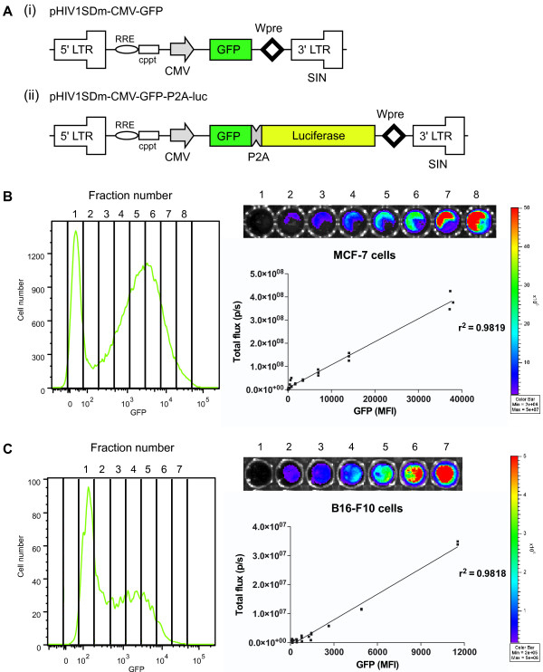
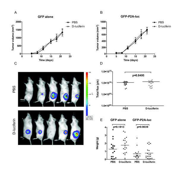
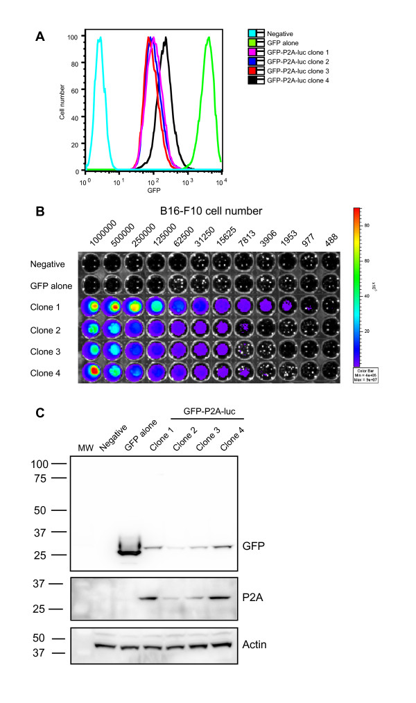
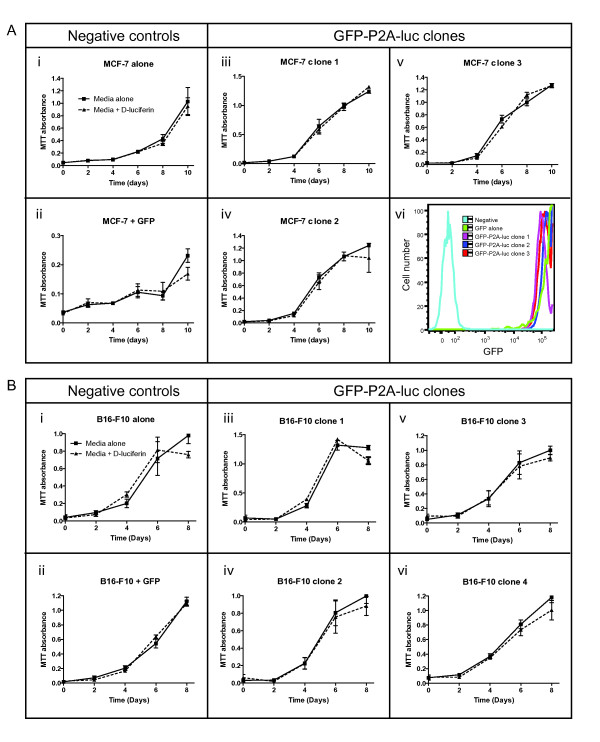
Live animal imaging is becoming an increasingly common technique for accurate and quantitative assessment of tumor burden over time. Bioluminescence imaging systems rely on a bioluminescent signal from tumor cells, typically generated from expression of the firefly luciferase gene. However, previous reports have suggested that either a high level of luciferase or the resultant light reaction produced upon addition of D-luciferin substrate can have a negative influence on tumor cell growth. To address this issue, we designed an expression vector that allows simultaneous fluorescence and luminescence imaging. Using fluorescence activated cell sorting (FACS), we generated clonal cell populations from a human breast cancer (MCF-7) and a mouse melanoma (B16-F10) cell line that stably expressed different levels of luciferase. We then compared the growth capabilities of these clones in vitro by MTT proliferation assay and in vivo by bioluminescence imaging of tumor growth in live mice. Surprisingly, we found that neither the amount of luciferase nor biophotonic activity was sufficient to inhibit tumor cell growth, in vitro or in vivo. These results suggest that luciferase toxicity is not a necessary consideration when designing bioluminescence experiments, and therefore our approach can be used to rapidly generate high levels of luciferase expression for sensitive imaging experiments.

摘要

活体动物成像是一种越来越常见的技术,可用于准确和定量评估肿瘤随时间的负担。生物发光成像系统依赖于肿瘤细胞的生物发光信号,通常是由萤火虫荧光素酶基因的表达产生的。然而,先前的报告表明,荧光素酶的高水平或加入 D-荧光素酶底物后产生的光反应可能对肿瘤细胞的生长产生负面影响。为了解决这个问题,我们设计了一个表达载体,允许同时进行荧光和发光成像。我们使用荧光激活细胞分选(FACS)从人乳腺癌(MCF-7)和小鼠黑色素瘤(B16-F10)细胞系中生成稳定表达不同水平荧光素酶的克隆细胞群。然后,我们通过 MTT 增殖测定法比较了这些克隆在体外的生长能力,并通过活体小鼠肿瘤生长的生物发光成像比较了它们在体内的生长能力。令人惊讶的是,我们发现无论是荧光素酶的数量还是生物光子活性都不足以抑制肿瘤细胞的生长,无论是在体外还是体内。这些结果表明,在设计生物发光实验时,荧光素酶毒性不是必需的考虑因素,因此我们的方法可用于快速生成高浓度的荧光素酶表达,以进行敏感的成像实验。

https://cdn.ncbi.nlm.nih.gov/pmc/blobs/71f3/3002927/950edf57970d/1476-4598-9-299-4.jpg
https://cdn.ncbi.nlm.nih.gov/pmc/blobs/71f3/3002927/9da2ea06cd3e/1476-4598-9-299-1.jpg
https://cdn.ncbi.nlm.nih.gov/pmc/blobs/71f3/3002927/f7ebf1f3492a/1476-4598-9-299-2.jpg
https://cdn.ncbi.nlm.nih.gov/pmc/blobs/71f3/3002927/83faab85dc06/1476-4598-9-299-3.jpg
https://cdn.ncbi.nlm.nih.gov/pmc/blobs/71f3/3002927/950edf57970d/1476-4598-9-299-4.jpg

相似文献

[1]
Luciferase expression and bioluminescence does not affect tumor cell growth in vitro or in vivo.

Mol Cancer. 2010-11-22

[2]
In vivo bioluminescence imaging of murine xenograft cancer models with a red-shifted thermostable luciferase.

Mol Imaging Biol. 2009-11-25

[3]
Rapid obtention of stable, bioluminescent tumor cell lines using a tCD2-luciferase chimeric construct.

BMC Biotechnol. 2011-3-24

[4]
The expression level of luciferase within tumour cells can alter tumour growth upon in vivo bioluminescence imaging.

Luminescence. 2007

[5]
In vitro validation of bioluminescent monitoring of disease progression and therapeutic response in leukaemia model animals.

Eur J Nucl Med Mol Imaging. 2006-5

[6]
Construction and validation of improved triple fusion reporter gene vectors for molecular imaging of living subjects.

Cancer Res. 2007-4-1

[7]
Evaluating Brightness and Spectral Properties of Click Beetle and Firefly Luciferases Using Luciferin Analogues: Identification of Preferred Pairings of Luciferase and Substrate for In Vivo Bioluminescence Imaging.

Mol Imaging Biol. 2020-12

[8]
The influence of hypoxia on bioluminescence in luciferase-transfected gliosarcoma tumor cells in vitro.

Photochem Photobiol Sci. 2008-6

[9]
Uptake kinetics and biodistribution of 14C-D-luciferin--a radiolabeled substrate for the firefly luciferase catalyzed bioluminescence reaction: impact on bioluminescence based reporter gene imaging.

Eur J Nucl Med Mol Imaging. 2008-12

[10]
Monitoring immediate-early gene expression through firefly luciferase imaging of HRS/J hairless mice.

BMC Physiol. 2003-8-19

引用本文的文献

[1]
Bioluminescent Imaging of Patient-Derived Brain Tumors in Live Mice.

Methods Mol Biol. 2025

[2]
In Vivo Bioluminescence Imaging of Tumor Progression in the Lewis Lung Carcinoma Orthotopic Mouse Model: A Comparison Between the Tail Vein Injection and Intranasal Instillation Methods.

Curr Protoc. 2024-12

[3]
Molecular Dynamics Simulation Combined with Neural Relationship Inference and Markov Model to Reveal the Relationship between Conformational Regulation and Bioluminescence Properties of Luciferase.

Molecules. 2024-8-26

[4]
PROTAC EZH2 degrader-1 overcomes the resistance of podophyllotoxin derivatives in refractory small cell lung cancer with leptomeningeal metastasis.

BMC Cancer. 2024-4-22

[5]
MALAT1 promotes FOXA1 degradation by competitively binding to miR-216a-5p and enhancing neuroendocrine differentiation in prostate cancer.

Transl Oncol. 2024-1

[6]
Carboxyl nanodiamonds inhibit melanoma tumor metastases by blocking cellular motility and invasiveness.

PNAS Nexus. 2023-11-28

[7]
Mismatch between Bioluminescence Imaging (BLI) and MRI When Evaluating Glioblastoma Growth: Lessons from a Study Where BLI Suggested "Regression" while MRI Showed "Progression".

Cancers (Basel). 2023-3-22

[8]
Pseudotyped Virus for Henipavirus.

Adv Exp Med Biol. 2023

[9]
Expression of tdTomato and luciferase in a murine lung cancer alters the growth and immune microenvironment of the tumor.

PLoS One. 2021

[10]
Portable bioluminescent platform for in vivo monitoring of biological processes in non-transgenic animals.

Nat Commun. 2021-5-11

本文引用的文献

[1]
The influence of hypoxia on bioluminescence in luciferase-transfected gliosarcoma tumor cells in vitro.

Photochem Photobiol Sci. 2008-6

[2]
Validity of bioluminescence measurements for noninvasive in vivo imaging of tumor load in small animals.

Biotechniques. 2007-7

[3]
The expression level of luciferase within tumour cells can alter tumour growth upon in vivo bioluminescence imaging.

Luminescence. 2007

[4]
Multimodality imaging of tumor xenografts and metastases in mice with combined small-animal PET, small-animal CT, and bioluminescence imaging.

J Nucl Med. 2007-2

[5]
Evaluation of firefly luciferase bioluminescence mediated photodynamic toxicity in cancer cells.

Mol Imaging Biol. 2006

[6]
Assessing tumor growth and distribution in a model of prostate cancer metastasis using bioluminescence imaging.

Clin Exp Metastasis. 2005

[7]
Noninvasive bioluminescence imaging of normal and spontaneously transformed prostate tissue in mice.

Cancer Res. 2006-5-1

[8]
Rapid analysis of T-cell selection in vivo using T cell-receptor retrogenic mice.

Nat Methods. 2006-3

[9]
Bioluminescence imaging.

Proc Am Thorac Soc. 2005

[10]
Optimisation of a multipartite human immunodeficiency virus based vector system; control of virus infectivity and large-scale production.

J Gene Med. 2005-11

文献AI研究员

20分钟写一篇综述,助力文献阅读效率提升50倍

立即体验

用中文搜PubMed

大模型驱动的PubMed中文搜索引擎

马上搜索

推荐工具

医学文档翻译智能文献检索