Hyakkoku Kana, Hamanaka Junya, Tsuruma Kazuhiro, Shimazawa Masamitsu, Hara Hideaki
Molecular Pharmacology, Department of Biofunctional Evaluation, Gifu Pharmaceutical University, 1-25-4 Daigaku-nishi, Gifu 501-1196, Japan.
Exp Transl Stroke Med. 2010 Nov 23;2(1):20. doi: 10.1186/2040-7378-2-20.
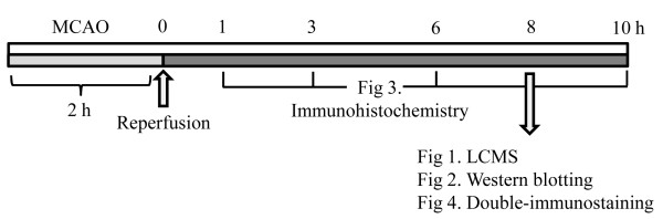
Several proteins are known to be markedly expressed in the brain during cerebral ischemia; however, the changes in protein profiles within the ischemic brain after an ischemic insult have not been fully elucidated. We studied the changes in the ischemic brain proteome after focal cerebral ischemia, induced by middle cerebral artery occlusion (MCAO) in mice.
LCMS-IT-TOF mass spectrometry was used to detect the changes in ischemic brain protein patterns after MCAO. We evaluated the protein expression detected in the ischemic area, by western blotting and immunohistochemistry.
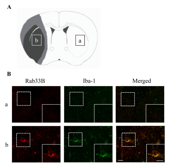
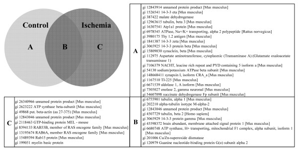
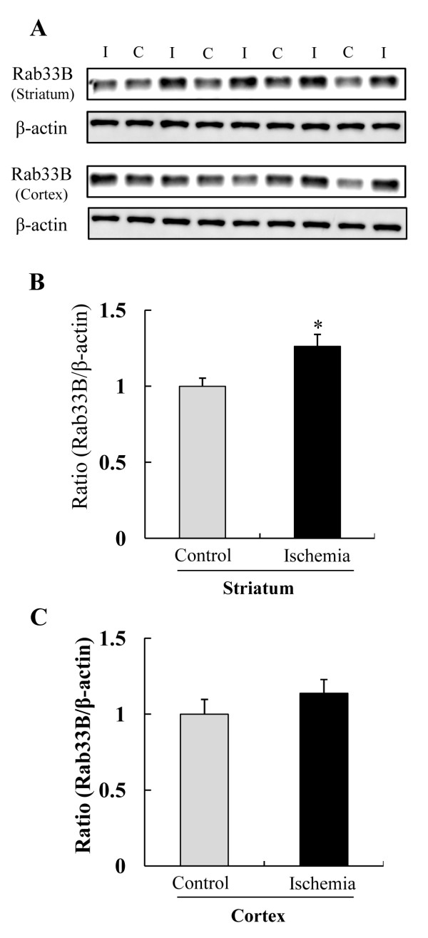
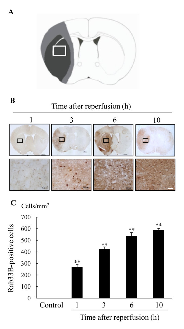
Nine unique proteins were identified from the ischemic area at 10 h after ischemic insult. Among these proteins, we focused on Rab33b, a member of RAS oncogene family and we found that Rab33b was up-regulated in the ischemic striatum and the number of Rab33B-positive cells increased in a time-dependent manner. Rab33B colocalized with Iba-1 positive microglia in the ischemic area.
These findings suggest that LCMS-IT-TOF is useful for identifying changes in proteins after cerebral ischemia and that Rab33B is partially related to the pathogenesis of transient cerebral ischemia in mice.
已知几种蛋白质在脑缺血期间在大脑中显著表达;然而,缺血性损伤后缺血性脑内蛋白质谱的变化尚未完全阐明。我们研究了小鼠大脑中动脉闭塞(MCAO)诱导的局灶性脑缺血后缺血性脑蛋白质组的变化。
采用液相色谱串联质谱仪(LCMS-IT-TOF)检测MCAO后缺血性脑蛋白质模式的变化。我们通过蛋白质印迹法和免疫组织化学评估在缺血区域检测到的蛋白质表达。
在缺血性损伤后10小时从缺血区域鉴定出9种独特的蛋白质。在这些蛋白质中,我们重点研究了RAS癌基因家族成员Rab33b,发现Rab33b在缺血纹状体中上调,且Rab33B阳性细胞数量呈时间依赖性增加。Rab33B在缺血区域与Iba-1阳性小胶质细胞共定位。
这些发现表明LCMS-IT-TOF可用于识别脑缺血后蛋白质的变化,且Rab33B与小鼠短暂性脑缺血的发病机制部分相关。