Ritzel Rodney M, Pan Sarah J, Verma Rajkumar, Wizeman John, Crapser Joshua, Patel Anita R, Lieberman Richard, Mohan Royce, McCullough Louise D
Neuroscience Department, University of Connecticut Health Center, Farmington, CT.
Neurology Department, University of Texas Health Science Center at Houston, Houston, TX.
Mol Vis. 2016 Jun 4;22:575-88. eCollection 2016.
The transient middle cerebral artery occlusion (MCAO) model of stroke is one of the most commonly used models to study focal cerebral ischemia. This procedure also results in the simultaneous occlusion of the ophthalmic artery that supplies the retina. Retinal cell death is seen days after reperfusion and leads to functional deficits; however, the mechanism responsible for this injury has not been investigated. Given that the eye may have a unique ocular immune response to an ischemic challenge, this study examined the inflammatory response to retinal ischemia in the MCAO model.
Young male C57B/6 mice were subjected to 90-min transient MCAO and were euthanized at several time points up to 7 days. Transcription of inflammatory cytokines was measured with quantitative real-time PCR, and immune cell activation (e.g., phagocytosis) and migration were assessed with ophthalmoscopy and flow cytometry.
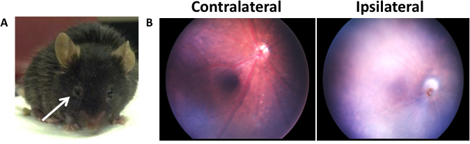
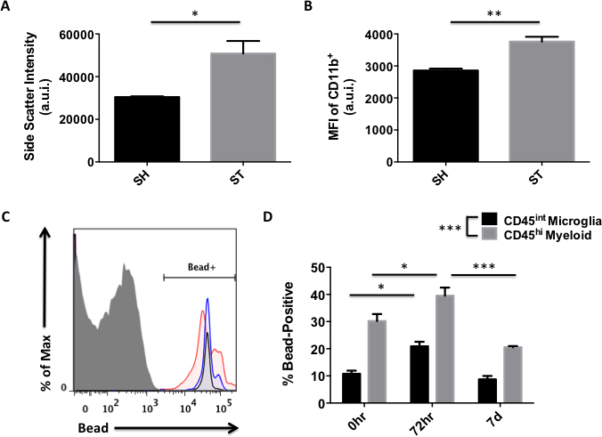
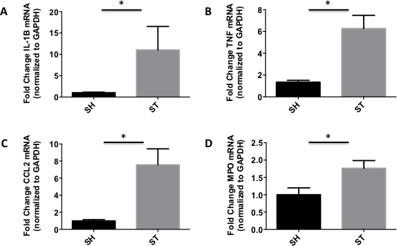
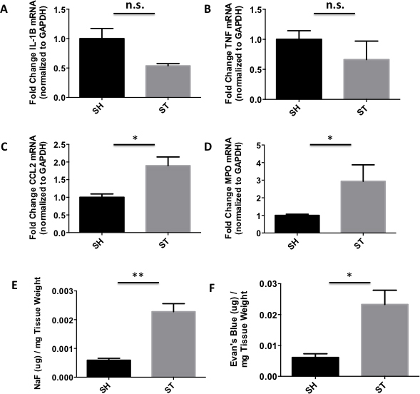
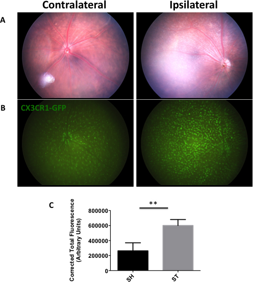
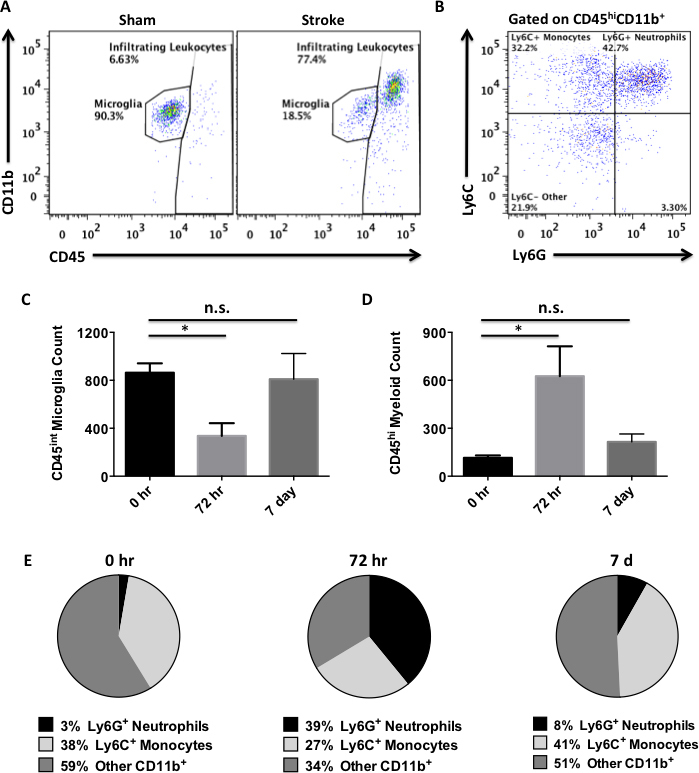
Observation of the affected eye revealed symptoms consistent with Horner's syndrome. Light ophthalmoscopy confirmed the reduced blood flow of the retinal arteries during occlusion. CX3CR1-GFP reporter mice were then employed to evaluate the extent of the ocular microglia and monocyte activation. A significant increase in green fluorescent protein (GFP)-positive macrophages was seen throughout the ischemic area compared to the sham and contralateral control eyes. RT-PCR revealed enhanced expression of the monocyte chemotactic molecule CCL2 early after reperfusion followed by a delayed increase in the proinflammatory cytokine TNF-α. Further analysis of peripheral leukocyte recruitment by flow cytometry determined that monocytes and neutrophils were the predominant immune cells to infiltrate at 72 h. A transient reduction in retinal microglia numbers was also observed, demonstrating the ischemic sensitivity of these cells. Blood-eye barrier permeability to small and large tracer molecules was increased by 72 h. Retinal microglia exhibited enhanced phagocytic activity following MCAO; however, infiltrating myeloid cells were significantly more efficient at phagocytizing material at all time points. Immune homeostasis in the affected eye was largely restored by 7 days.
This work demonstrates that there is a robust inflammatory response in the eye following MCAO, which may contribute to a worsening of retinal injury and visual impairment. These results mirror what has been observed in the brain after MCAO, suggesting a conserved inflammatory signaling response to ischemia in the central nervous system. Imaging of the eye may therefore serve as a useful non-invasive prognostic indicator of brain injury after MCAO. Future studies are needed to determine whether this inflammatory response is a potential target for therapeutic manipulation in retinal ischemia.
短暂性大脑中动脉闭塞(MCAO)卒中模型是研究局灶性脑缺血最常用的模型之一。该操作还会导致供应视网膜的眼动脉同时闭塞。视网膜细胞死亡在再灌注数天后出现,并导致功能缺陷;然而,造成这种损伤的机制尚未得到研究。鉴于眼睛可能对缺血性刺激有独特的眼部免疫反应,本研究在MCAO模型中检测了对视网膜缺血的炎症反应。
将年轻雄性C57B/6小鼠进行90分钟的短暂性MCAO,并在长达7天的多个时间点实施安乐死。用定量实时PCR测量炎症细胞因子的转录,并通过检眼镜检查和流式细胞术评估免疫细胞的激活(如吞噬作用)和迁移。
对患眼的观察发现了与霍纳综合征一致的症状。光学检眼镜检查证实闭塞期间视网膜动脉血流减少。然后使用CX3CR1-GFP报告基因小鼠来评估眼部小胶质细胞和单核细胞的激活程度。与假手术组和对侧对照眼相比,在整个缺血区域观察到绿色荧光蛋白(GFP)阳性巨噬细胞显著增加。逆转录聚合酶链反应(RT-PCR)显示再灌注后早期单核细胞趋化分子CCL2表达增强,随后促炎细胞因子TNF-α延迟增加。通过流式细胞术对外周白细胞募集的进一步分析确定,单核细胞和中性粒细胞是在72小时时浸润的主要免疫细胞。还观察到视网膜小胶质细胞数量短暂减少,证明了这些细胞对缺血的敏感性。到72小时时,血眼屏障对大小示踪分子的通透性增加。MCAO后视网膜小胶质细胞表现出增强的吞噬活性;然而,在所有时间点,浸润的髓样细胞吞噬物质的效率明显更高。到7天时,患眼中的免疫稳态基本恢复。
这项研究表明,MCAO后眼睛会出现强烈的炎症反应,这可能导致视网膜损伤和视力损害加重。这些结果与MCAO后在大脑中观察到的情况相似,表明中枢神经系统对缺血存在保守的炎症信号反应。因此,眼部成像可能作为MCAO后脑损伤有用的非侵入性预后指标。未来需要开展研究来确定这种炎症反应是否是视网膜缺血治疗干预的潜在靶点。