Biswas Sondip K, Lee Jai Eun, Brako Lawrence, Jiang Jean X, Lo Woo-Kuen
Department of Neurobiology, Morehouse School of Medicine, Atlanta, GA 30310, USA.
Mol Vis. 2010 Nov 9;16:2328-41.
Ball-and-sockets and protrusions are specialized interlocking membrane domains between lens fibers of all species studied. Ball-and-sockets and protrusions are similar in their shape, size, and surface morphology, and are traditionally believed to play a key role in maintaining fiber-to-fiber stability. Here, we evaluate the hypothesis that ball-and-sockets and protrusions possess important structural and functional differences during fiber cell differentiation and maturation.
Intact lenses of leghorn chickens (E7 days to P62 weeks old) and rhesus monkeys (1.5-20 years old) were studied with SEM, freeze-fracture TEM, freeze-fracture immunogold labeling (FRIL), and filipin cytochemistry for membrane cholesterol detection.
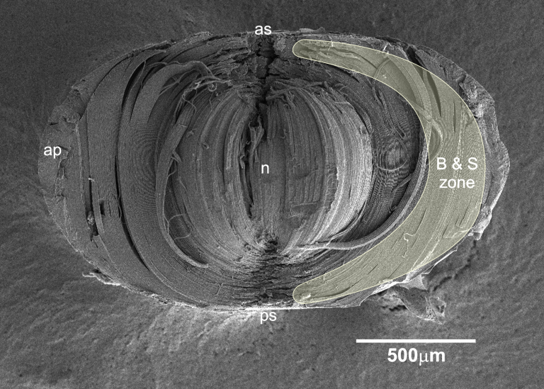
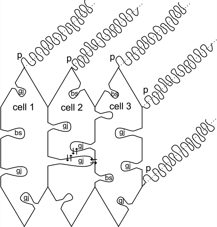
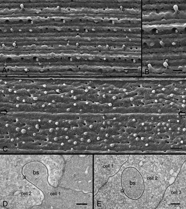
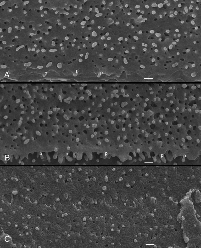
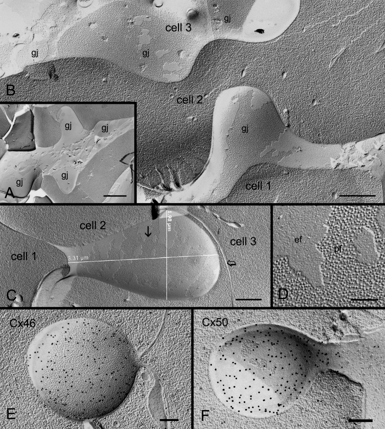
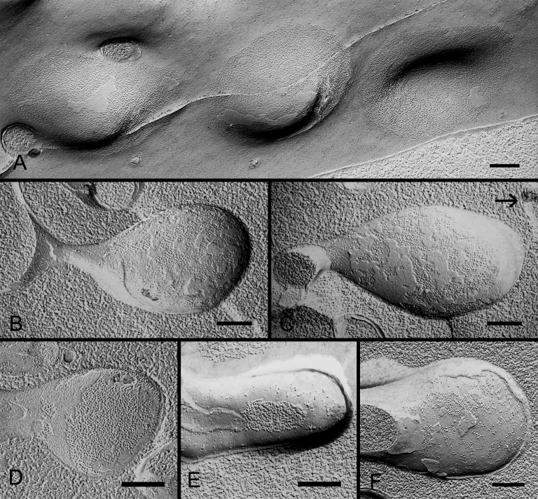
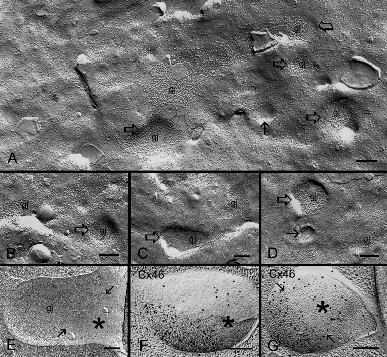
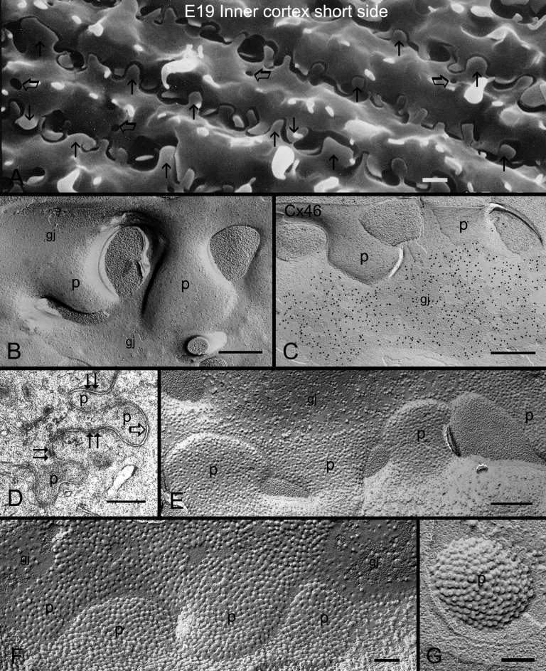
SEM showed that ball-and-sockets were distributed along the long and short sides of hexagonal fiber cells, whereas protrusions were located along the cell corners, from superficial to deep cortical regions in both chicken and monkey lenses. Importantly, by freeze-fracture TEM, we discovered the selective association of gap junctions with all ball-and-sockets examined, but not with protrusions, in both species. In the embryonic chicken lens (E18), the abundant distribution of ball-and-socket gap junctions was regularly found in an approximate zone extending at least 300 μm deep from the equatorial surface of the superficial cortical fibers. Many ball-and-socket gap junctions often protruded deeply into neighboring cells. However, in the mature fibers of monkey lenses, several ball-and-sockets exhibited only partial occupancy of gap junctions with disorganized connexons, possibly due to degradation of gap junctions during fiber maturation and aging. FRIL analysis confirmed that both connexin46 (Cx46) and connexin50 (Cx50) antibodies specifically labeled ball-and-socket gap junctions, but not protrusions. Furthermore, filipin cytochemistry revealed that the ball-and-socket gap junctions contained different amounts of cholesterol (i.e., cholesterol-rich versus cholesterol-free) as seen with the filipin-cholesterol-complexes (FCC) in different cortical regions during maturation. In contrast, the protrusions contained consistently high cholesterol amounts (i.e., 402 FCCs/μm2 membrane) which were approximately two times greater than that of the cholesterol-rich gap junctions (i.e., 188 FCCs/μm2 membrane) found in ball-and-sockets.
Gap junctions are regularly associated with all ball-and-sockets examined in metabolically active young cortical fibers, but not with protrusions, in both chicken and monkey lenses. Since these unique gap junctions often protrude deeply into neighboring cells to increase membrane surface areas, they may significantly facilitate cell-to-cell communication between young cortical fiber cells. In particular, the large number of ball-and-socket gap junctions found near the equatorial region may effectively facilitate the flow of outward current toward the equatorial surface for internal circulation of ions in the lens. In contrast, a consistent distribution of high concentrations of cholesterol in protrusions would make the protrusion membrane less deformable and would be more suitable for maintaining fiber-to-fiber stability during visual accommodation. Thus, the ball-and-sockets and protrusions are two structurally and functionally distinct membrane domains in the lens.
球窝和突起是所有已研究物种的晶状体纤维之间特化的互锁膜结构域。球窝和突起在形状、大小和表面形态上相似,传统上认为它们在维持纤维间稳定性方面起关键作用。在此,我们评估这样一种假说,即球窝和突起在纤维细胞分化和成熟过程中具有重要的结构和功能差异。
使用扫描电子显微镜(SEM)、冷冻断裂透射电子显微镜(freeze - fracture TEM)、冷冻断裂免疫金标记(FRIL)以及用于膜胆固醇检测的制霉菌素细胞化学方法,对来亨鸡(胚胎期7天至出生后62周龄)和恒河猴(1.5 - 20岁)的完整晶状体进行研究。
SEM显示,球窝沿六边形纤维细胞的长边和短边分布,而突起位于细胞角,在鸡和猴的晶状体中从浅皮质区域到深皮质区域均有。重要的是,通过冷冻断裂TEM,我们发现在这两个物种中,缝隙连接与所有检测的球窝选择性相关,但与突起无关。在胚胎期鸡的晶状体(E18)中,在从浅皮质纤维赤道表面至少深入300μm的大致区域内,经常能发现丰富分布的球窝缝隙连接。许多球窝缝隙连接常常深深突入相邻细胞。然而,在猴晶状体的成熟纤维中,一些球窝仅显示缝隙连接的部分占据且连接子排列紊乱,这可能是由于缝隙连接在纤维成熟和老化过程中发生了降解。FRIL分析证实,连接蛋白46(Cx46)和连接蛋白50(Cx50)抗体特异性标记球窝缝隙连接,而不标记突起。此外,制霉菌素细胞化学显示,随着晶状体成熟,在不同皮质区域,球窝缝隙连接含有不同量的胆固醇(即富含胆固醇与不含胆固醇),这可通过制霉菌素 - 胆固醇复合物(FCC)观察到。相比之下,突起含有始终较高的胆固醇量(即402个FCCs/μm²膜),约为球窝中富含胆固醇的缝隙连接(即188个FCCs/μm²膜)的两倍。
在鸡和猴的晶状体中,缝隙连接与代谢活跃的年轻皮质纤维中所有检测的球窝有规律地相关,但与突起无关。由于这些独特的缝隙连接常常深深突入相邻细胞以增加膜表面积,它们可能显著促进年轻皮质纤维细胞之间的细胞间通讯。特别是,在赤道区域附近发现的大量球窝缝隙连接可能有效地促进外向电流流向赤道表面,以实现晶状体中离子的内部循环。相反,突起中高浓度胆固醇的一致分布会使突起膜的可变形性降低,更适合在视觉调节过程中维持纤维间稳定性。因此,球窝和突起是晶状体中两个结构和功能不同的膜结构域。