Center for Musculoskeletal Surgery, Julius Wolff Institute Berlin, Charité – Universitaetsmedizin Berlin, Germany.
Acta Orthop. 2011 Feb;82(1):102-11. doi: 10.3109/17453674.2010.539498. Epub 2010 Dec 13.
Animal models of skeletal muscle injury should be thoroughly described and should mimic the clinical situation. We established a model of a critical size crush injury of the soleus muscle in rats. The aim was to describe the time course of skeletal muscle regeneration using mechanical, histological, and magnetic resonance (MR) tomographic methods.
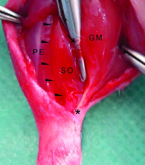
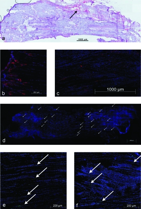
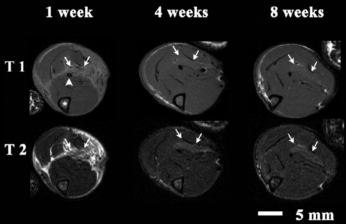
Left soleus muscles of 36 Sprague-Dawley rats were crushed in situ in a standardized manner. We scanned the lower legs of 6 animals by 7-tesla MR one week, 4 weeks, and 8 weeks after trauma. Regeneration was evaluated at these times by in vivo measurement of muscle contraction forces after fast-twitch and tetanic stimulation (groups 1W, 4W, 8W; 6 per group). Histological and immunohistological analysis was performed and the amount of fibrosis within the injured muscles was determined histomorphologically.
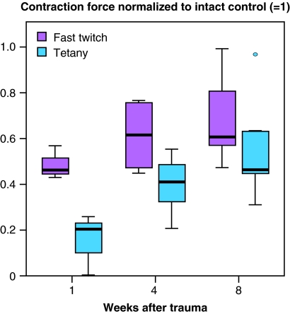
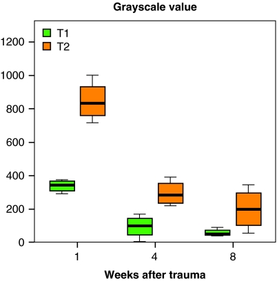
MR signals of the traumatized soleus muscles showed a clear time course concerning microstructure and T1 and T2 signal intensity. Newly developed neural endplates and myotendinous junctions could be seen in the injured zones of the soleus. Tetanic force increased continuously, starting at 23% (SD 4) of the control side (p < 0.001) 1 week after trauma and recovering to 55% (SD 23) after 8 weeks. Fibrotic tissue occupied 40% (SD 4) of the traumatized muscles after the first week, decreased to approximately 25% after 4 weeks, and remained at this value until 8 weeks.
At both the functional level and the morphological level, skeletal muscle regeneration follows a distinct time course. Our trauma model allows investigation of muscle regeneration after a standardized injury to muscle fibers.
骨骼肌损伤的动物模型应进行全面描述,并模拟临床情况。我们建立了大鼠比目鱼肌严重挤压伤的模型。目的是使用机械、组织学和磁共振(MR)断层扫描方法描述骨骼肌再生的时间过程。
采用标准化方法原位挤压 36 只 Sprague-Dawley 大鼠的左比目鱼肌。创伤后 1 周、4 周和 8 周,我们使用 7 特斯拉 MR 对 6 只动物的小腿进行扫描。在这些时间点,通过快速抽搐和强直刺激后活体测量肌肉收缩力(每组 6 只,分别为 1W、4W、8W 组)来评估再生情况。进行组织学和免疫组织化学分析,并通过组织形态学确定损伤肌肉内的纤维化程度。
受损伤的比目鱼肌的 MR 信号在微观结构以及 T1 和 T2 信号强度方面呈现出明确的时间过程。在比目鱼肌的损伤区域可以看到新形成的神经终板和肌腱连接。强直力从创伤后 1 周开始持续增加,最初为健侧的 23%(标准差 4%)(p < 0.001),8 周后恢复至 55%(标准差 23%)。创伤后第 1 周,纤维化组织占损伤肌肉的 40%(标准差 4%),4 周后降至约 25%,直至 8 周保持不变。
在功能和形态水平上,骨骼肌再生都遵循明确的时间过程。我们的创伤模型允许在标准化的肌纤维损伤后研究肌肉再生。