Laboratoire de Biologie des APS, EA 3533, PRES Clermont Université, Université Blaise Pascal, 24 Avenue des Landais, BP 80026, 63177 Aubière Cedex, France.
Lipids Health Dis. 2010 Dec 9;9:140. doi: 10.1186/1476-511X-9-140.
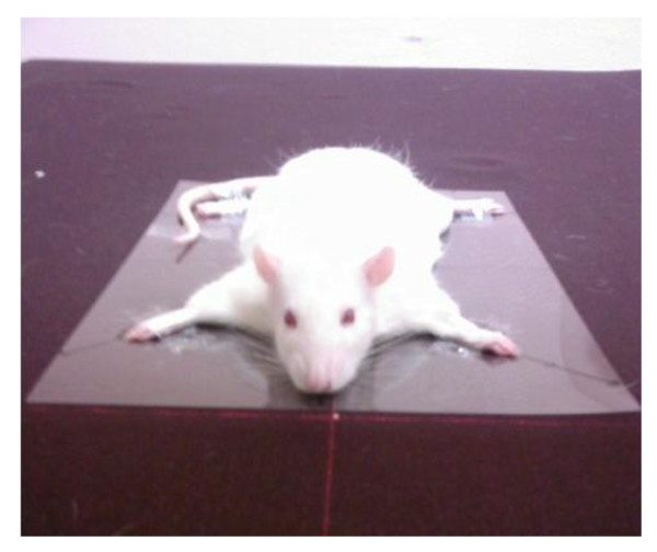
Because abdominal obesity is predisposed to various metabolic disorders, it is of major importance to assess and track the changes with time of this specific fat mass. The main issue for clinicians or researchers is to use techniques for assessing abdominal fat deposition and its accumulation or changes over time, without sacrificing of experimental subjects. In the rat, techniques to investigate in-vivo visceral fat mass are lacking. The purpose of the study was to validate indirect Dual-energy X-ray Absorptiometry technique and abdominal circumference measurement as tools to predict visceral adipose tissue in rats.Forty-three Wistar male rats from different body weight, fat mass and ages were included in the study. Visceral fat mass was assessed by weighing the total perirenal and peri-epididymal adipose tissues after dissection. Statistical methods were used to discriminate the best region of interest allowing the in-vivo measure of Central Fat Mass by DXA. Abdominal circumference was measured at the same time as the DXA scan.
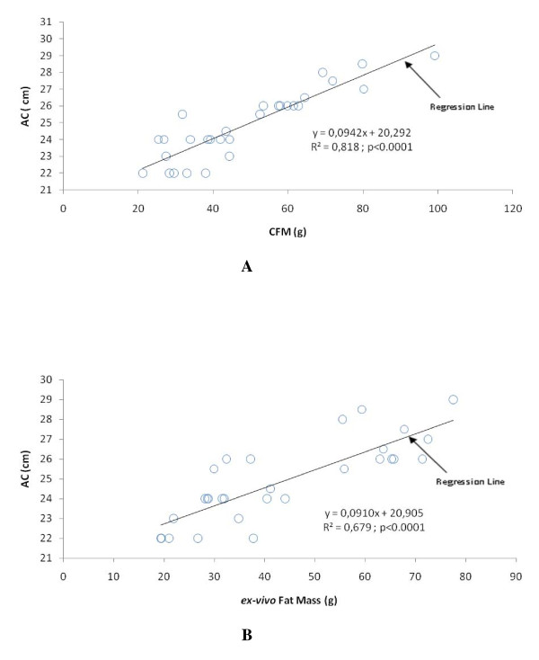
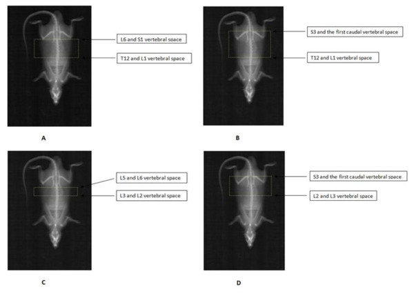
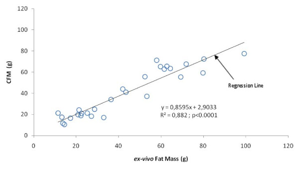
A region of interest including Central Fat Mass from the whole body DXA scan (extending from L2 to L5 vertebrae), correlated strongly with ex-vivo Fat Mass (r = 0.94, p < 0.001). Abdominal circumference correlated significantly with ex-vivo Fat Mass (r = 0.82, p < 0.001) and Central Fat Mass (0.90, p < 0.001) in the whole group of rats. When dividing the whole group into lean and fat rats, correlations remained significant between Central Fat Mass and ex-vivo Fat Mass but disappeared for the lean group between abdominal circumference and ex-vivo Fat Mass.
This study validates the Central Fat Mass determined by DXA as a non-sacrificial technique to assess visceral fat for in-vivo investigations in rats. The abdominal circumference measure appears useful in studying overweight or obese rats. These two techniques could be convenient tools in follow-up and longitudinal studies.
由于腹型肥胖易导致各种代谢紊乱,因此评估和跟踪特定脂肪量随时间的变化非常重要。临床医生或研究人员的主要问题是使用评估腹部脂肪沉积及其随时间的积累或变化的技术,而不牺牲实验对象。在大鼠中,缺乏研究内脏脂肪量的技术。本研究旨在验证间接双能 X 射线吸收法技术和腹围测量作为预测大鼠内脏脂肪组织的工具。
研究纳入了 43 只来自不同体重、体脂和年龄的雄性 Wistar 大鼠。解剖后称重总肾周和附睾周脂肪组织来评估内脏脂肪量。使用统计方法来区分最佳感兴趣区域,以便通过 DXA 进行体内中央脂肪量测量。同时在 DXA 扫描时测量腹围。
包括整个 DXA 扫描的中央脂肪量感兴趣区域(从 L2 到 L5 椎骨延伸),与体外脂肪量高度相关(r = 0.94,p < 0.001)。腹围与整个大鼠组的体外脂肪量(r = 0.82,p < 0.001)和中央脂肪量(0.90,p < 0.001)显著相关。当将整个组分为瘦大鼠和胖大鼠时,中央脂肪量与体外脂肪量之间的相关性仍然显著,但在瘦大鼠组中,腹围与体外脂肪量之间的相关性消失。
本研究验证了 DXA 确定的中央脂肪量是一种非牺牲性技术,可用于评估大鼠体内内脏脂肪。腹围测量在研究超重或肥胖大鼠时很有用。这两种技术可能是随访和纵向研究的便利工具。