Zhu Hong-Yuan, Riau A K, Beuerman R W
Singapore Eye Research Institute, Yong Loo Lin School of Medicine, National University of Singapore, Singapore.
Mol Vis. 2010 Oct 31;16:2215-24.
The conjunctival epithelium is a continuous sheet of cells with regional characteristics that appear to be similar. This study was designed to investigate the distribution and levels of expression of a subset of microfilament regulators in the forniceal, palpebral, and bulbar conjunctival epithelia.
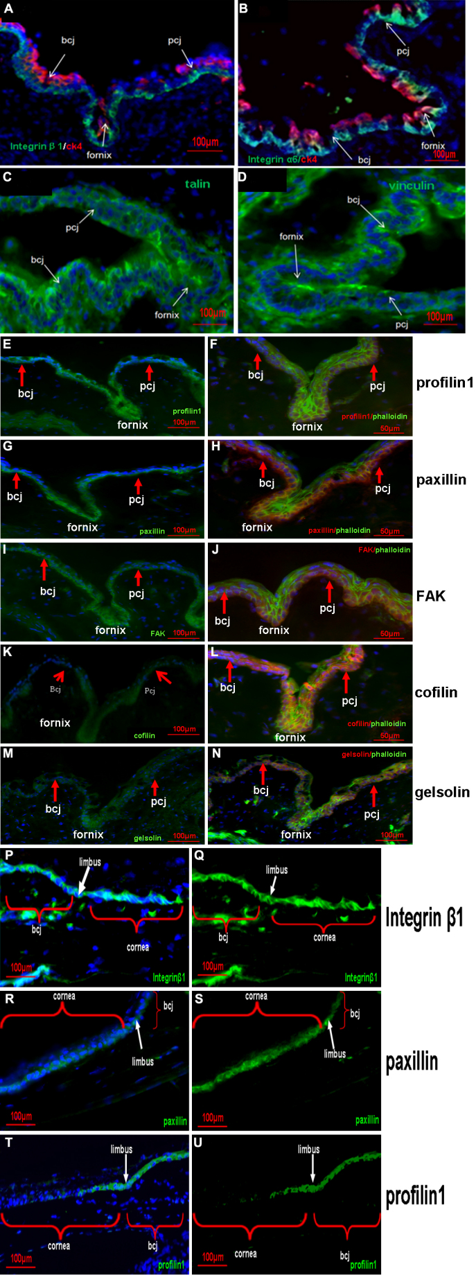
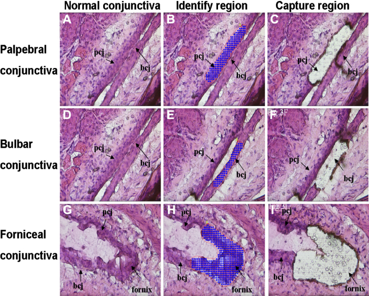
Balb/C mice were used. The localizations of paxillin, focal adhesion kinase, vinculin, talin1, cofilin, profilin, gelsolin, integrin β1, and integrin α6 were studied with the use of cross-sectional immunofluorescent staining. For a detailed cellular analysis, positioning and ablation with the laser microbeam (PALM) Combi System was used to obtain forniceal, bulbar, and palpebral conjunctival epithelia for expression comparison with the use of western blot analysis and quantitative real-time polymerase chain reaction.
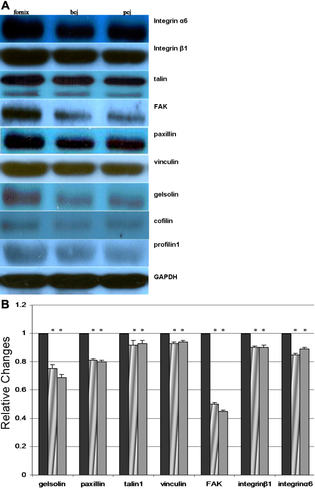
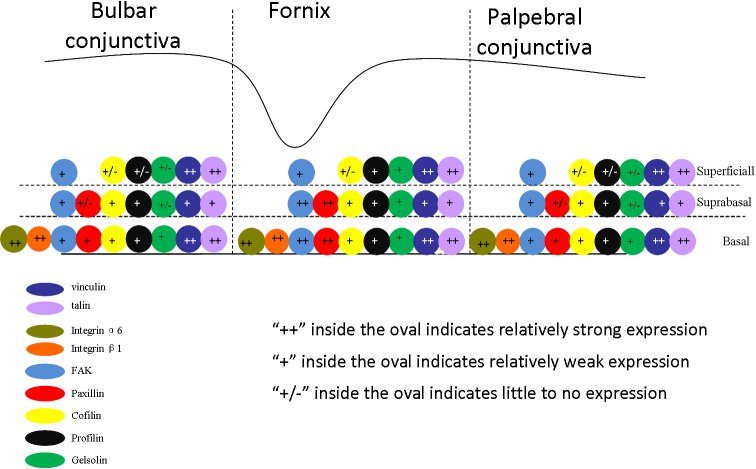
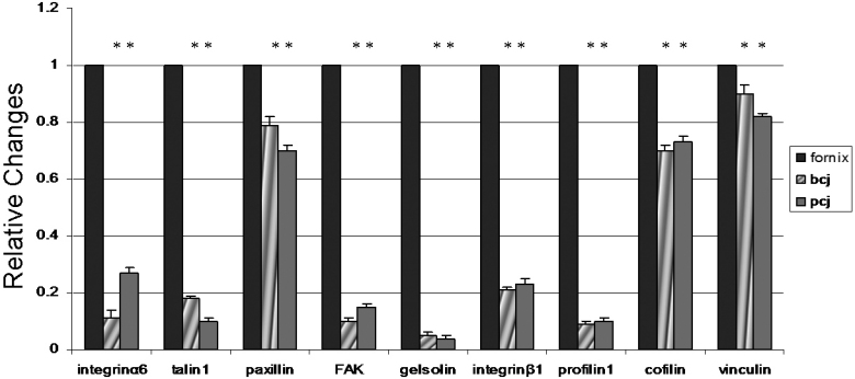
Immunostaining showed that focal adhesion kinase, cofilin, profilin, gelsolin, talin1, and vinculin were expressed in all layers of the forniceal, palpebral, and bulbar conjunctival epithelia. Paxillin, integrin β1, and α6 was found to be located in the basal cell layer in all three of these areas. Quantitative real-time polymerase chain reaction showed that the transcript levels of these microfilament regulators in the forniceal conjunctivae were higher than those levels found in the bulbar and palpebral conjunctivae. Western blot analysis confirmed the differential expression levels of these microfilament regulators in the forniceal, bulbar, and palpebral conjunctivae.
Differences in the levels of microfilament regulators in the forniceal, bulbar, and palpebral conjunctivae suggest different modes of interaction with their microenvironment and within cell layers.
结膜上皮是一层连续的细胞,具有看似相似的区域特征。本研究旨在调查微丝调节因子亚群在穹窿部、睑部和球结膜上皮中的分布及表达水平。
使用Balb/C小鼠。采用横断面免疫荧光染色研究桩蛋白、粘着斑激酶、纽蛋白、踝蛋白1、丝切蛋白、前纤维蛋白、凝溶胶蛋白、整合素β1和整合素α6的定位。为进行详细的细胞分析,使用激光微束(PALM)Combi系统进行定位和切除,以获取穹窿部、球部和睑部结膜上皮,用于通过蛋白质免疫印迹分析和定量实时聚合酶链反应进行表达比较。
免疫染色显示,粘着斑激酶、丝切蛋白、前纤维蛋白、凝溶胶蛋白、踝蛋白1和纽蛋白在穹窿部、睑部和球结膜上皮的所有层中均有表达。发现桩蛋白、整合素β1和α6位于所有这三个区域的基底细胞层。定量实时聚合酶链反应显示,这些微丝调节因子在穹窿结膜中的转录水平高于在球结膜和睑结膜中的水平。蛋白质免疫印迹分析证实了这些微丝调节因子在穹窿部、球部和睑部结膜中的差异表达水平。
穹窿部、球部和睑部结膜中微丝调节因子水平的差异表明它们与微环境以及细胞层内相互作用的方式不同。