Laboratory of Clinical Biochemistry and Molecular Biology, Department of Laboratory Medicine and Molecular Diagnostics, University Hospital of Pisa Pisa, Italy.
Front Syst Neurosci. 2010 Nov 22;4:152. doi: 10.3389/fnsys.2010.00152. eCollection 2010.
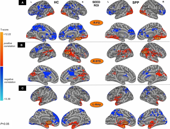
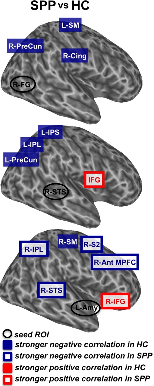
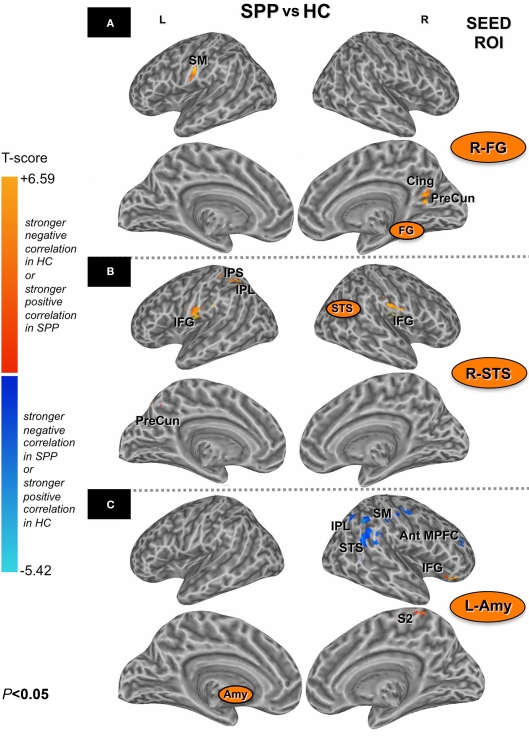
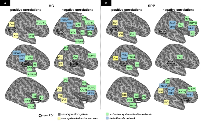
Recently, a differential recruitment of brain areas throughout the distributed neural system for face perception has been found in social phobic patients as compared to healthy control subjects. These functional abnormalities in social phobic patients extend beyond emotion-related brain areas, such as the amygdala, to include cortical networks that modulate attention and process other facial features, and they are also associated with an alteration of the task-related activation/deactivation trade-off. Functional connectivity is becoming a powerful tool to examine how components of large-scale distributed neural systems are coupled together while performing a specific function. This study was designed to determine whether functional connectivity networks among brain regions within the distributed system for face perception also would differ between social phobic patients and healthy controls. Data were obtained from eight social phobic patients and seven healthy controls by using functional magnetic resonance imaging. Our findings indicated that social phobic patients and healthy controls have different patterns of functional connectivity across brain regions within both the core and the extended systems for face perception and the default mode network. To our knowledge, this is the first study that shows that functional connectivity during brain response to socially relevant stimuli differs between social phobic patients and healthy controls. These results expand our previous findings and indicate that brain functional changes in social phobic patients are not restricted to a single specific brain structure, but rather involve a mis-communication among different sensory and emotional processing brain areas.
最近,与健康对照组相比,社交恐惧症患者在分布式神经系统中整个大脑区域对面孔感知的差异招募已经被发现。社交恐惧症患者的这些功能异常不仅延伸到与情绪相关的大脑区域(如杏仁核),还包括调节注意力和处理其他面部特征的皮质网络,并且它们还与任务相关的激活/失活权衡的改变有关。功能连接正成为一种强大的工具,可以检查在执行特定功能时,大型分布式神经系统的组件如何相互耦合。这项研究旨在确定在执行特定功能时,面孔感知分布式系统内的大脑区域之间的功能连接网络是否也会在社交恐惧症患者和健康对照组之间存在差异。通过使用功能磁共振成像,从八名社交恐惧症患者和七名健康对照者中获取数据。我们的研究结果表明,社交恐惧症患者和健康对照组在面孔感知的核心系统和扩展系统以及默认模式网络内的大脑区域之间具有不同的功能连接模式。据我们所知,这是第一项表明在对社交相关刺激的大脑反应中,社交恐惧症患者和健康对照组之间的功能连接存在差异的研究。这些结果扩展了我们之前的发现,并表明社交恐惧症患者的大脑功能变化不仅限于单个特定的大脑结构,而是涉及不同感觉和情绪处理大脑区域之间的错误通信。